 뒤로
뒤로Chapter 1: An Introduction to Anatomy and Physiology – Study Notes
스터디 가이드 - 스마트 노트
 자료에 맞춘 맞춤형 노트, 핵심 정의, 예시, 맥락을 확장해 제공합니다.
Introduction to Anatomy & Physiology
Overview of Anatomy and Physiology
Anatomy and physiology are foundational sciences in understanding the structure and function of the human body. Anatomy focuses on the physical structures, while physiology explores how these structures function to sustain life. The integration of these disciplines is essential for comprehending how the body operates as a whole.
Anatomy: The study of internal and external body structures and their relationships.
Physiology: The study of how living organisms perform vital functions.
Principle of Complementarity: Structure and function are closely related; specific functions are performed by specific structures.
Specialties in Anatomy and Physiology
Types of Anatomy
Gross (Macroscopic) Anatomy: Study of large structures visible to the naked eye.
Regional Anatomy: Focuses on specific areas of the body.
Systemic Anatomy: Examines organ systems.
Microscopic Anatomy: Study of structures requiring magnification.
Cytology: Study of cells.
Histology: Study of tissues.
Types of Physiology
Cell Physiology: Functions of cells and their chemical processes.
Organ Physiology: Functions of specific organs.
Systemic Physiology: Functions of organ systems.
Pathological Physiology: Effects of diseases on organs or systems.
Levels of Organization in the Human Body
Hierarchical Structure
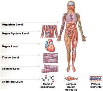
The human body is organized into six major levels, from the simplest chemical components to the complex organism.
Chemical Level: Atoms and molecules (e.g., water, proteins).
Cellular Level: Cells, the smallest living units.
Tissue Level: Groups of similar cells performing specific functions. Four basic tissue types: epithelial, connective, muscle, and neural.
Organ Level: Two or more tissues working together (e.g., heart).
Organ System Level: Groups of organs performing related functions (e.g., cardiovascular system).
Organism Level: The complete living individual.


Origins and Standardization of Anatomical Terms
Importance of Terminology
Standardized anatomical terminology ensures clear communication among healthcare professionals. Many terms are derived from Latin or Greek, providing consistency and precision in describing body structures and functions.
Anatomical Position and Body Regions
Standard Anatomical Position
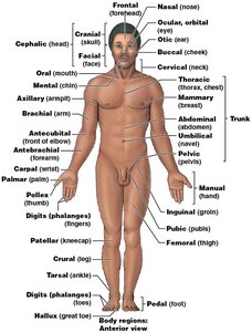
The anatomical position is the reference posture for describing locations and directions on the human body: standing upright, facing forward, arms at the sides, palms facing forward, and feet together.
Supine: Lying face up.
Prone: Lying face down.
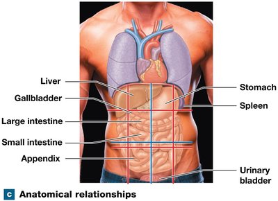
Abdominopelvic Quadrants and Regions
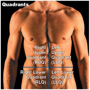
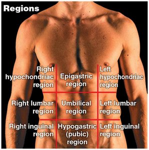
The abdominopelvic area is divided for clinical and anatomical reference:
Quadrants: Right Upper (RUQ), Left Upper (LUQ), Right Lower (RLQ), Left Lower (LLQ).
Regions: Nine regions including epigastric, umbilical, hypogastric, and others for detailed localization.
Directional Terms and Sectional Anatomy
Directional References
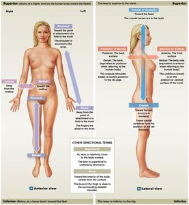
Directional terms describe the positions of structures relative to each other:
Superior: Toward the head.
Inferior: Toward the feet.
Anterior (Ventral): Toward the front.
Posterior (Dorsal): Toward the back.
Medial: Toward the midline.
Lateral: Away from the midline.
Proximal: Closer to the point of attachment.
Distal: Farther from the point of attachment.
Superficial: Toward the surface.
Deep: Away from the surface.
Sectional Planes
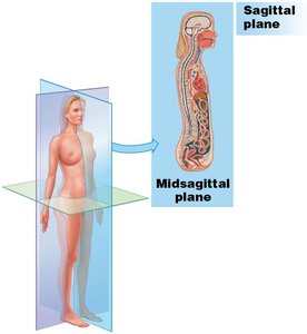
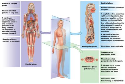
Sectional anatomy involves slicing the body along specific planes to view internal structures:
Frontal (Coronal) Plane: Divides the body into anterior and posterior portions.
Sagittal Plane: Divides the body into left and right portions. The midsagittal plane is exactly in the middle; parasagittal is offset.
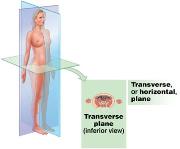
Transverse (Horizontal) Plane: Divides the body into superior and inferior portions.
Medical Imaging Techniques
Overview of Imaging Modalities
Medical imaging allows visualization of internal structures for diagnosis and research:
MRI (Magnetic Resonance Imaging): Uses magnetic fields and radio waves; does not expose patients to radiation.
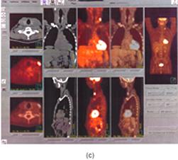
PET (Positron Emission Tomography): Uses radioactive substances to visualize metabolic activity.
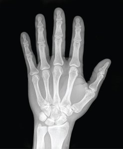
X-ray: Uses high-energy electromagnetic radiation to view dense structures like bones.
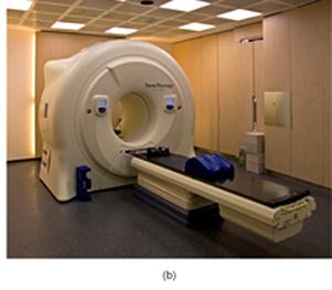
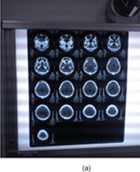
CT (Computed Tomography): Combines multiple X-rays for detailed cross-sectional images; higher radiation dose than standard X-rays.
Body Cavities and Membranes
Major Body Cavities
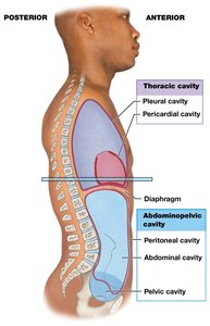
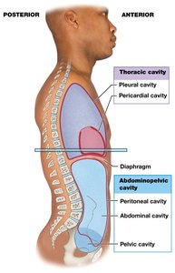
Body cavities are closed, fluid-filled spaces that house and protect internal organs (viscera). They allow organs to change shape and size and are lined by serous membranes.
Thoracic Cavity: Contains pleural cavities (lungs), pericardial cavity (heart), and mediastinum.
Abdominopelvic Cavity: Contains abdominal and pelvic cavities, as well as the peritoneal cavity.
Serous Membranes
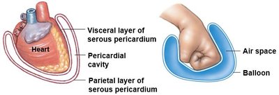
Parietal Layer: Lines the cavity wall.
Visceral Layer: Covers the organ.
Serous Fluid: Reduces friction between layers.
Homeostasis
Definition and Importance
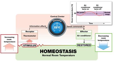
Homeostasis is the maintenance of a stable internal environment despite external changes. It is essential for normal physiological function and survival.
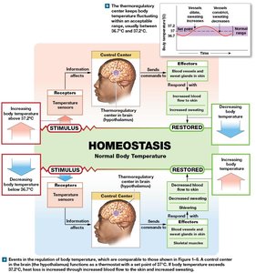
Receptor: Detects changes (stimuli).
Control Center: Processes information and sends commands.
Effector: Carries out the response to restore balance.
Set Point: The ideal value for a physiological variable.
Feedback Mechanisms
Negative Feedback
Negative feedback opposes deviations from the set point, maintaining variables within a normal range. Most homeostatic mechanisms in the body use negative feedback.
Example: Regulation of body temperature, blood glucose levels.
Positive Feedback
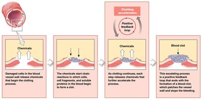
Positive feedback amplifies changes, moving the variable further from the set point. It is used for processes that must be rapidly completed.
Example: Blood clotting, childbirth contractions.
Summary Table: Levels of Organization
Level | Description | Example |
|---|---|---|
Chemical | Atoms and molecules | Water, proteins |
Cellular | Smallest living units | Muscle cell |
Tissue | Groups of similar cells | Muscle tissue |
Organ | Two or more tissues | Heart |
Organ System | Group of organs | Cardiovascular system |
Organism | Complete individual | Human body |
Additional info: The scientific method is used in clinical diagnosis, involving observation, hypothesis formation, and experimental testing. Signs are objective indicators of disease, while symptoms are subjective experiences reported by the patient.
