 뒤로
뒤로Chapter 1: An Introduction to Anatomy and Physiology – Structured Study Notes
스터디 가이드 - 스마트 노트
 자료에 맞춘 맞춤형 노트, 핵심 정의, 예시, 맥락을 확장해 제공합니다.
Introduction to Anatomy & Physiology
Overview
Anatomy and physiology are foundational sciences in understanding the structure and function of the human body. Anatomy focuses on the physical structures, while physiology explores how these structures operate and interact. The relationship between anatomy and physiology is integral, as the form of a structure is closely tied to its function.
Anatomy: Study of body structures, their composition, and location.
Physiology: Study of body functions, both individual and cooperative.
Relationship: The physiological function of a system depends on its anatomy, and anatomy is designed to fit its physiological function.
Specialties of Anatomy and Physiology
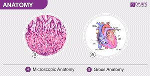
Gross Anatomy: Examines large, visible structures (e.g., surface anatomy).
Microscopic Anatomy: Examines cells and molecules (cytology and histology).
Human Physiology: Includes cell physiology, organ physiology, systemic physiology, and pathological physiology.
Levels of Organization in Organisms
Hierarchical Structure
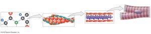
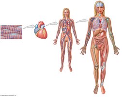
The human body is organized into several levels, from the simplest chemical components to the complex organism. Each level builds upon the previous, contributing to the overall function and structure of the body.
Chemical Level: Atoms and molecules form the basis of all matter.
Cellular Level: Cells are the smallest living units, composed of molecules.
Tissue Level: Groups of cells working together to perform specific functions.
Organ Level: Organs are made of multiple tissue types working together.
Organ System Level: Organ systems consist of interacting organs.
Organism Level: The complete living individual.
The Organ Systems
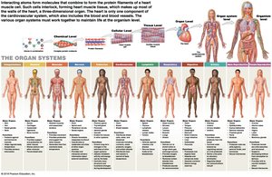
Humans have 11 major organ systems, each with specific organs and functions. These systems work together to maintain homeostasis and support life.
Integumentary System: Skin, hair, nails; protection and temperature regulation.
Skeletal System: Bones, cartilage; support, protection, mineral storage.
Muscular System: Muscles, tendons; movement, heat production.
Nervous System: Brain, spinal cord, nerves; coordination and sensory information.
Endocrine System: Glands; regulation of metabolism and development.
Cardiovascular System: Heart, blood vessels; transport of nutrients and waste.
Lymphatic System: Spleen, lymph nodes; defense against infection.
Respiratory System: Lungs, airways; gas exchange.
Digestive System: Stomach, intestines; food processing and absorption.
Urinary System: Kidneys, bladder; waste excretion and fluid balance.
Reproductive System: Ovaries/testes; production of sex cells and hormones.
Medical Terminology
Structure of Medical Terms
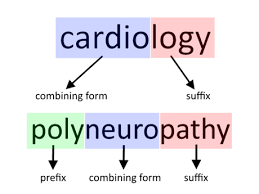
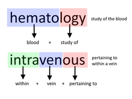
Medical terminology is essential for clear communication in health and disease. Terms are constructed from word roots, prefixes, suffixes, and combining forms.
Word Root: Basic meaning of the term.
Prefix: Added to the beginning to modify meaning.
Suffix: Added to the end to modify meaning.
Combining Form: Connects word parts for pronunciation.
Anatomical Terminology
Surface Anatomy and Anatomical Landmarks
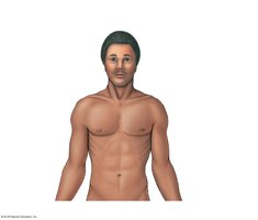
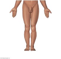
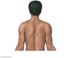
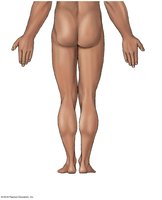
Surface anatomy involves locating structures on or near the body surface. The standard anatomical position is used as a reference: hands at sides, palms forward.
Landmarks: Frontal, nasal, orbital, cranial, thoracic, abdominal, etc.
Anterior and Posterior Views: Used to describe locations and regions.
Abdominopelvic Quadrants and Regions
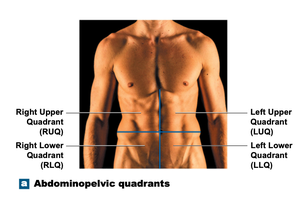
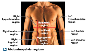
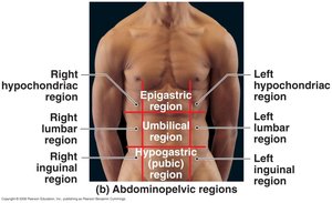
The abdominopelvic area is divided for clinical and anatomical purposes. Quadrants are used by medical personnel, while regions are used by anatomists.
Quadrants: Right Upper (RUQ), Left Upper (LUQ), Right Lower (RLQ), Left Lower (LLQ).
Regions: Nine regions including epigastric, umbilical, hypogastric, lumbar, inguinal, hypochondriac.
Directional Terms
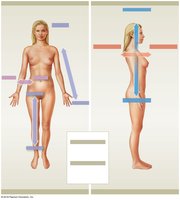
Directional terms are used to describe the location of structures relative to each other.
Superior: Above; toward the head.
Inferior: Below; toward the feet.
Anterior (Ventral): Front side.
Posterior (Dorsal): Back side.
Medial: Toward the midline.
Lateral: Away from the midline.
Proximal: Toward the point of attachment.
Distal: Away from the point of attachment.
Superficial: Near the surface.
Deep: Farther from the surface.
Sectional Anatomy and Planes
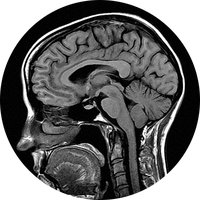
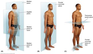
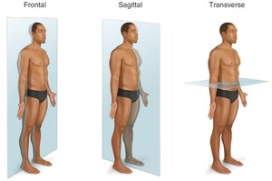
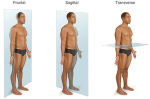
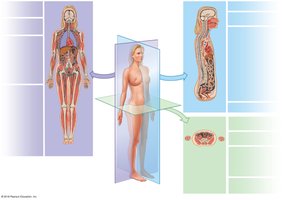
Sectional anatomy involves slicing the body to visualize internal structures. Common planes include frontal (coronal), sagittal, and transverse.
Frontal (Coronal) Plane: Divides body into anterior and posterior portions.
Sagittal Plane: Divides body into left and right portions (midsagittal and parasagittal).
Transverse Plane: Divides body into superior and inferior portions.
Body Cavities
Functions and Structure
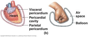
Body cavities are closed, fluid-filled spaces lined by serous membranes. They protect internal organs and allow changes in organ size and shape.
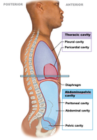
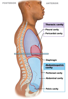
Ventral Body Cavity: Divided by the diaphragm into thoracic and abdominopelvic cavities.
Serous Membrane: Parietal layer lines cavity, visceral layer covers organ, serous fluid reduces friction.
Thoracic and Abdominopelvic Cavities
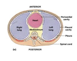
Thoracic Cavity: Contains pleural cavities (lungs), pericardial cavity (heart), and mediastinum (connective tissue and major vessels).
Abdominopelvic Cavity: Contains abdominal cavity (digestive organs), pelvic cavity (reproductive organs, rectum, bladder), and peritoneal cavity.
Homeostasis
Concept and Regulation
Homeostasis is the maintenance of a stable internal environment despite external fluctuations. All body systems contribute to this balance, responding to changes to keep variables within normal ranges.
Dynamic Equilibrium: Continual adaptation to maintain balance.
Homeostatic Regulation: Adjustment of physiological systems via autoregulation (local) and extrinsic regulation (nervous/endocrine systems).
Homeostatic Regulatory Mechanism
Receptor: Detects stimulus.
Control Center: Processes information and sends instructions.
Effector: Carries out instructions to restore balance.
Negative and Positive Feedback
Negative Feedback
Negative feedback mechanisms counteract changes, bringing the body back to homeostasis. Most physiological processes use negative feedback to maintain stability.
Example: Regulation of body temperature.
Positive Feedback
Positive feedback amplifies changes, moving the body away from homeostasis. It is used for rapid completion of processes, such as blood clotting and childbirth.
Example: Blood clotting cascade.
Systems Integration and Disease
Systems Integration
Physiological systems work together to maintain homeostasis. Failure of integration leads to disease, as seen in diabetes mellitus, where insufficient insulin disrupts blood glucose regulation.
Dynamic Equilibrium: Balance between opposing forces.
Disease: Imbalance leads to loss of function and cell death.
Example: Diabetes mellitus results from insufficient insulin production, causing elevated blood sugar and loss of homeostasis.
