 뒤로
뒤로Chapter 1: Introduction to Anatomy and Physiology – Structured Study Notes
스터디 가이드 - 스마트 노트
 자료에 맞춘 맞춤형 노트, 핵심 정의, 예시, 맥락을 확장해 제공합니다.
An Introduction to Anatomy and Physiology
Definition and Scope of Anatomy & Physiology
Anatomy and physiology are foundational sciences in understanding the structure and function of the human body. Anatomy describes the physical structures, their composition, location, and associated features. Physiology focuses on the functions of these anatomical structures, both individually and in cooperation with other systems.
Anatomy: Study of body structures, including what they are made of, where they are located, and their associated structures.
Physiology: Study of the functions of anatomical structures, including individual and cooperative functions.
Branches of Anatomy
Gross (Macroscopic) Anatomy: Examines large, visible structures.
Surface Anatomy: Study of exterior features.
Regional Anatomy: Study of specific body areas.
Sectional Anatomy: Study of cross sections.
Systemic Anatomy: Study of organ systems.
Clinical Anatomy: Medical specialties.
Developmental Anatomy: From conception to adulthood, including embryology.
Microscopic Anatomy: Examines cells and molecules (cytology and histology).
Branches of Physiology
Cell Physiology: Functions of cells.
Organ Physiology: Functions of specific organs.
Systemic Physiology: Functions of organ systems.
Pathological Physiology: Effects of diseases on organs or systems.
Levels of Organization
Hierarchical Structure of the Human Body
The human body is organized into a hierarchy of structural levels, each building upon the previous. Understanding these levels is essential for grasping how the body functions as a whole.
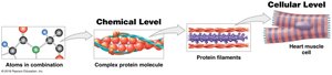
Chemical Level: Atoms and molecules form the basis of all matter.
Cellular Level: Cells are the smallest living units in the body.
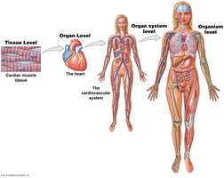
Tissue Level: Groups of cells working together to perform specific functions.
Organ Level: Organs are made of two or more tissues working together.
Organ System Level: Groups of interacting organs.
Organism Level: An individual life form.
Example: The heart muscle cell (cellular level) forms cardiac muscle tissue (tissue level), which makes up the heart (organ level), part of the cardiovascular system (organ system level), contributing to the organism (human body).
Organ Systems Overview
Major Organ Systems and Their Functions
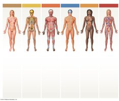
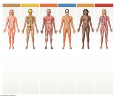
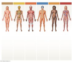
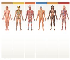
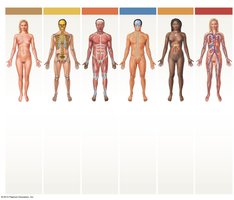
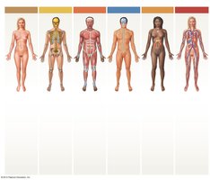
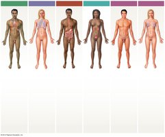
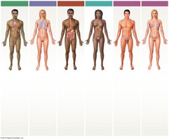
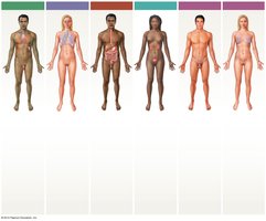
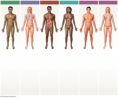
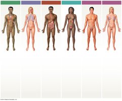
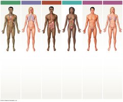
The human body contains 11 major organ systems, each with specific organs and functions. These systems work together to maintain health and homeostasis.
Integumentary System: Skin, hair, sweat glands, nails. Protects against environmental hazards, regulates temperature, provides sensory information.
Skeletal System: Bones, cartilages, ligaments, bone marrow. Provides support, protection, stores minerals, forms blood cells.
Muscular System: Skeletal muscles, tendons. Provides movement, protection, support, generates heat.
Nervous System: Brain, spinal cord, nerves, sense organs. Directs responses, coordinates systems, interprets sensory information.
Endocrine System: Glands (pituitary, thyroid, adrenal), pancreas, gonads. Directs long-term changes, adjusts metabolism, controls development.
Cardiovascular System: Heart, blood, blood vessels. Distributes blood, nutrients, waste, oxygen, carbon dioxide, heat.
Lymphatic System: Spleen, thymus, lymph nodes, vessels, tonsils. Defends against infection, returns fluids to bloodstream.
Respiratory System: Nasal cavities, sinuses, larynx, trachea, bronchi, lungs, alveoli. Delivers air, provides oxygen, removes carbon dioxide, produces sounds.
Digestive System: Teeth, tongue, pharynx, esophagus, stomach, intestines, liver, gallbladder, pancreas. Processes food, absorbs nutrients and water, stores energy.
Urinary System: Kidneys, ureters, bladder, urethra. Excretes waste, regulates water and ions, stores urine, maintains pH.
Reproductive System: Male: testes, ducts, glands, penis, scrotum. Female: ovaries, tubes, uterus, vagina, labia, clitoris, mammary glands. Produces sex cells, hormones, supports embryo, provides milk.
Anatomical Terminology
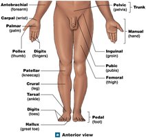
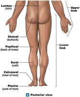
Surface and Anatomical Landmarks
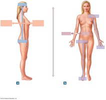
Anatomical terminology is essential for accurately describing locations and relationships of body structures. Surface anatomy involves locating structures on or near the body surface using landmarks and reference positions.
Anatomical Position: Hands at sides, palms forward.
Supine: Lying face up.
Prone: Lying face down.
Anatomical Regions and Directions
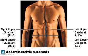
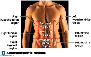
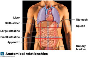
The abdominopelvic region is divided into quadrants and regions for clinical and descriptive purposes. Directional terms are used to describe the location of structures relative to each other.
Abdominopelvic Quadrants: Right Upper (RUQ), Left Upper (LUQ), Right Lower (RLQ), Left Lower (LLQ).
Abdominopelvic Regions: More precise, includes epigastric, umbilical, hypogastric, lumbar, inguinal, hypochondriac regions.
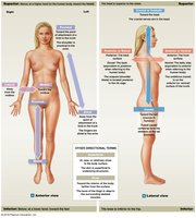
Directional Terms: Superior, inferior, anterior (ventral), posterior (dorsal), medial, lateral, proximal, distal.
Sectional Anatomy and Diagnostic Imaging
Sectional Planes
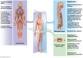
Sectional anatomy involves slicing through the body to visualize internal structures. This is important in medical imaging and diagnosis.
Frontal (Coronal) Plane: Divides body into anterior and posterior portions.
Sagittal Plane: Divides body into left and right portions. Midsagittal is exactly in the middle; parasagittal is offset.
Transverse Plane: Divides body into superior and inferior portions.
Diagnostic Imaging Techniques
Modern imaging techniques allow visualization of internal structures for diagnosis and treatment.
X-rays: High energy radiation, radiopaque tissues appear white.
CT Scan: Computed tomography, shows 3D relationships and soft tissues.
MRI: Magnetic resonance imaging, shows soft tissues in detail.
PET Scan: Assesses metabolic and physiological activity.
Ultrasound: Uses high-frequency sound waves, produces echograms.
Spiral Scan: 3D imaging, less radiation than CT.
DSA: Digital subtraction angiography, monitors blood flow with high contrast.
Body Cavities
Functions and Structure of Body Cavities
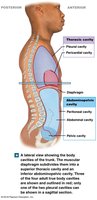
Body cavities protect internal organs and allow changes in size and shape. The ventral body cavity is divided by the diaphragm into thoracic and abdominopelvic cavities, each containing specific organs and lined by serous membranes.
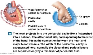
Thoracic Cavity: Contains lungs (pleural cavities), heart (pericardial cavity), mediastinum.
Abdominopelvic Cavity: Contains digestive, urinary, and reproductive organs.
Serous Membrane: Parietal layer lines cavity, visceral layer covers organ.
Homeostasis
Concept and Mechanisms of Homeostasis
Homeostasis is the maintenance of a stable internal environment. All body systems contribute to this balance, responding to changes to keep variables within normal ranges.
Autoregulation (Intrinsic): Automatic response in a cell, tissue, or organ.
Extrinsic Regulation: Controlled by nervous and endocrine systems.
Homeostatic Regulatory Mechanism
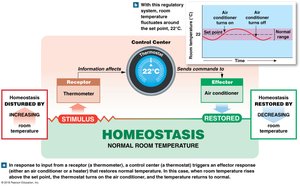
A homeostatic mechanism consists of a receptor (detects stimulus), control center (processes information), and effector (carries out instructions). This limits fluctuations and maintains a set point.
Negative and Positive Feedback
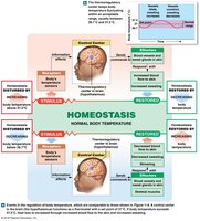
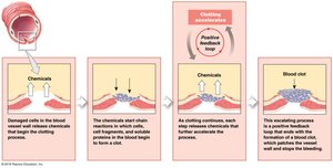
Feedback mechanisms regulate homeostasis. Negative feedback opposes changes, restoring balance. Positive feedback amplifies changes, often used to complete processes quickly.
Negative Feedback: Response negates the stimulus, restoring homeostasis.
Positive Feedback: Response amplifies the stimulus, moving away from homeostasis.
Systems Integration and Equilibrium
Physiological systems work together to maintain equilibrium. Failure to maintain homeostasis results in disease.
