 뒤로
뒤로Chapter 1: Introduction to Anatomy & Physiology – Structured Study Notes
스터디 가이드 - 스마트 노트
 자료에 맞춘 맞춤형 노트, 핵심 정의, 예시, 맥락을 확장해 제공합니다.
Introduction to Anatomy & Physiology
Characteristics of Living Things
Living organisms exhibit several fundamental characteristics that distinguish them from non-living matter. These characteristics are essential for maintaining life and adapting to environmental changes.
Organization: Each organism maintains a distinct boundary separating it from its environment. Internal conditions are often very different from external conditions.
Responsiveness: The ability to respond to changes in the immediate environment.
Irritability: The capacity to detect and react to stimuli.
Adaptability: Adjustments made to cope with environmental changes.
Growth & Differentiation: Increase in size and number of cells; cells grow and then become specialized types.

Anatomy & Physiology: Definitions and Branches
Anatomy and physiology are closely related fields that study the structure and function of living organisms. Understanding both is crucial for comprehending how the human body operates.
Anatomy: The study of internal and external structures of the body and the physical relationships among body parts.
Gross Anatomy (Macroscopic Anatomy): Study of large structures visible with the naked eye.
Microscopic Anatomy: Study of structures that cannot be seen without magnification.
Cytology: Study of cell structure.
Histology: Study of tissues (groups of specialized cells).
Physiology: Study of how organisms perform their vital functions.
Form Follows Function: The structure of a body part is determined by its function.

Levels of Organization in the Human Body
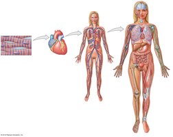
Chemical and Cellular Levels
The human body is organized into hierarchical levels, starting from the simplest chemical components to the complex cellular structures.
Chemical Level: Atoms combine to form molecules, such as proteins and nucleic acids.
Cellular Level: Molecules form organelles, which make up cells—the basic unit of life.
Example: Heart muscle cells contain protein filaments and complex protein molecules.

Tissue, Organ, and Organ System Levels
Cells group together to form tissues, which then combine to create organs. Organs work together in organ systems to perform vital functions.
Tissue Level: Groups of similar cells performing a specific function (e.g., cardiac muscle tissue).
Organ Level: Organs are made up of different tissues working together (e.g., the heart).
Organ System Level: Organ systems consist of multiple organs that coordinate to perform complex functions (e.g., cardiovascular system).
Organism Level: The highest level, representing the complete living individual.
The Organ Systems of the Human Body
Overview of Major Organ Systems
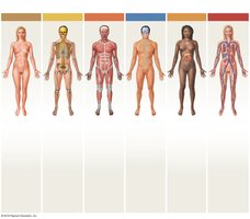
The human body contains several organ systems, each with specific organs and functions. These systems work together to maintain homeostasis and support life.
Organ System | Major Organs | Functions |
|---|---|---|
Integumentary | Skin, hair, sweat glands, nails | Protection, temperature regulation, sensory information |
Skeletal | Bones, cartilages, ligaments, bone marrow | Support, protection, mineral storage, blood cell formation |
Muscular | Skeletal muscles, tendons | Movement, heat generation, protection |
Nervous | Brain, spinal cord, nerves, sense organs | Immediate responses, coordination, sensory interpretation |
Endocrine | Pituitary, thyroid, pancreas, adrenal glands, gonads | Long-term regulation, metabolism, development |
Cardiovascular | Heart, blood, blood vessels | Transport, heat distribution |
Lymphatic | Spleen, thymus, lymphatic vessels, nodes, tonsils | Defense, fluid return |
Respiratory | Nasal cavities, sinuses, larynx, trachea, bronchi, lungs, alveoli | Gas exchange, sound production |
Digestive | Teeth, tongue, pharynx, esophagus, stomach, intestines, liver, gallbladder, pancreas | Food processing, absorption, water conservation |
Urinary | Kidneys, ureters, bladder, urethra | Waste excretion, water balance, ion regulation |
Male Reproductive | Testes, epididymides, ductus deferentia, seminal vesicles, prostate, penis, scrotum | Sperm production, hormones, sexual intercourse |
Female Reproductive | Ovaries, uterine tubes, uterus, vagina, labia, clitoris, mammary glands | Oocyte production, hormones, embryo support, milk production, sexual intercourse |

Homeostasis and Feedback Mechanisms
Homeostasis: Definition and Importance
Homeostasis is the maintenance of a stable internal environment despite external changes. It is essential for the survival and proper functioning of organisms.
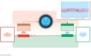
Set Point: The ideal value for a physiological parameter (e.g., body temperature).
Normal Range: The range of values around the set point that are considered healthy.
Control Center: Receives information from receptors and directs effectors to restore balance.
Receptor: Detects changes in the environment.
Effector: Responds to commands from the control center to restore homeostasis.
Negative Feedback
Negative feedback is a regulatory mechanism in which a deviation from the set point triggers a response that counteracts the change, restoring homeostasis.
Example: Regulation of body temperature. If body temperature rises above 37.2ºC, the hypothalamus triggers increased blood flow to the skin and sweating to cool the body. If temperature falls below 36.7ºC, shivering and decreased blood flow to the skin help raise temperature.
Key Steps: Stimulus → Receptor → Control Center → Effector → Response → Restoration of homeostasis
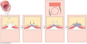
Positive Feedback
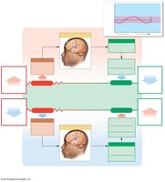
Positive feedback amplifies a change instead of reversing it. This mechanism is less common but important in certain physiological processes.
Example: Blood clotting. Damaged cells release chemicals that initiate clotting. Each step releases more chemicals, accelerating the process until the clot forms and bleeding stops.
Key Steps: Stimulus → Chain reaction → Amplification → Completion of process
Anatomical Terminology and Landmarks
Body Regions and Landmarks
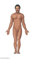
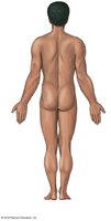
Anatomical terminology provides precise descriptions of locations and regions on the body, which is essential for communication in healthcare and science.
Anterior (front) view: Includes regions such as frontal, nasal, ocular, thoracic, abdominal, pelvic, brachial, femoral, etc.
Posterior (back) view: Includes regions such as cephalic, acromial, dorsal, lumbar, gluteal, popliteal, sural, calcaneal, plantar, etc.
Abdominopelvic Quadrants and Regions
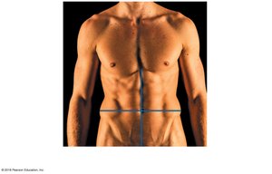
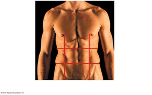
The abdominopelvic area is divided into quadrants and regions to aid in diagnosis and description of symptoms.
Quadrants: Right Upper (RUQ), Right Lower (RLQ), Left Upper (LUQ), Left Lower (LLQ)
Regions: Right/Left hypochondriac, epigastric, umbilical, hypogastric (pubic), right/left lumbar, right/left inguinal
Directional References
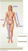
Directional terms describe the positions of structures relative to each other. These terms are fundamental for anatomical orientation.
Superior: Above; toward the head
Inferior: Below; toward the feet
Anterior (Ventral): Front surface
Posterior (Dorsal): Back surface
Medial: Toward the midline
Lateral: Away from the midline
Proximal: Toward the point of attachment
Distal: Away from the point of attachment
Superficial: Near the body surface
Deep: Toward the interior of the body

Sectional Planes
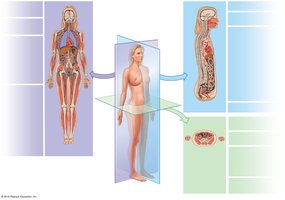
Sectional planes are used to describe cuts or views of the body, which are important in medical imaging and anatomical study.
Frontal (Coronal) Plane: Separates anterior and posterior portions.
Sagittal Plane: Separates right and left portions. Midsagittal passes through the midline; parasagittal misses the midline.
Transverse (Horizontal) Plane: Separates superior and inferior portions.
Body Cavities and Their Subdivisions
Major Body Cavities
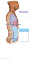
The trunk of the body contains several major cavities that house vital organs. These cavities are separated by membranes and the diaphragm.
Thoracic Cavity: Contains pleural cavities (lungs) and pericardial cavity (heart).
Abdominopelvic Cavity: Contains abdominal cavity (digestive organs) and pelvic cavity (reproductive and urinary organs).
Diaphragm: Muscular partition separating thoracic and abdominopelvic cavities.
Serous Membranes and Cavity Relationships
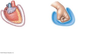
Serous membranes line body cavities and cover organs, providing lubrication and reducing friction.
Visceral Layer: Covers the organ.
Parietal Layer: Lines the cavity wall.
Pericardial Cavity: Surrounds the heart, with a thin layer of fluid between visceral and parietal layers.
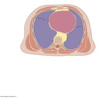
Transverse Section of Thoracic Cavity
A transverse (cross-sectional) view of the thoracic cavity shows the central location of the pericardial cavity and its relationship to the pleural cavities.
Mediastinum: Central compartment containing the heart, major vessels, and other structures.
Pleural Cavities: Each contains a lung.
Clinical Relevance: Transverse views are standard in diagnostic imaging.
Additional info: Some explanations and context were expanded for clarity and completeness, based on standard academic knowledge in anatomy and physiology.
