 뒤로
뒤로Chapter 1: The Human Body—An Orientation (Anatomy & Physiology Study Notes)
스터디 가이드 - 스마트 노트
 자료에 맞춘 맞춤형 노트, 핵심 정의, 예시, 맥락을 확장해 제공합니다.
Introduction to Anatomy & Physiology
Overview of Anatomy and Physiology
Anatomy and physiology are foundational sciences in understanding the human body. Anatomy is the study of the structure of the human body, while physiology focuses on the functions of body parts and systems. These disciplines are essential for medical professionals, scientists, and artists who require knowledge of the human form and function.
Anatomy: Examines the physical structures of organisms.
Physiology: Explores how anatomical structures operate and interact.
Studied by: Scientists, healthcare professionals, kinesthesiologists, therapists, and artists.
Historical figures: Leonardo da Vinci (artist and anatomist), Herophilus of Alexandria ("father of anatomy"), Andreas Vesalius (author of De Humani Corporis Fabrica).
Subdisciplines and Branches of Anatomy
Major Subdisciplines
Anatomy is divided into several subdisciplines, each focusing on different levels of organization or methods of study.
Gross Anatomy: Study of structures visible to the naked eye.
Regional Anatomy: Focuses on specific areas (e.g., head, chest).
Systemic Anatomy: Studies body systems (e.g., skeletal, muscular).
Surface Anatomy: Examines external features as landmarks for deeper structures.
Microscopic Anatomy (Histology): Study of tissues and cells using microscopes.
Developmental Anatomy: Explores structural changes throughout life.
Embryology: Focuses on development before birth.
Pathological Anatomy: Studies structural changes caused by disease.
Radiographic Anatomy: Uses imaging techniques to view internal structures.
Functional Morphology: Investigates the relationship between structure and function.
The Hierarchy of Structural Organization
Levels of Organization
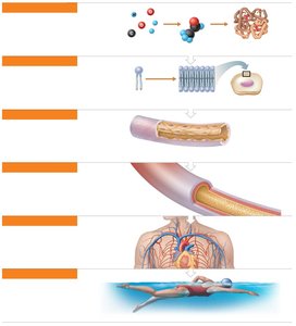
The human body is organized into a hierarchy of structural levels, each building upon the previous one. Understanding these levels is crucial for grasping how the body functions as a whole.
Chemical Level: Atoms combine to form molecules (e.g., carbohydrates, proteins).
Cellular Level: Cells and their organelles are the basic units of life.
Tissue Level: Groups of similar cells perform common functions.
Organ Level: Organs are made of multiple tissue types working together.
Organ System Level: Groups of organs perform related functions.
Organismal Level: The complete living individual, resulting from the integration of all systems.
Major Organ Systems of the Human Body
Overview of Organ Systems
The human body consists of several organ systems, each with specific functions essential for survival and homeostasis.
Integumentary System: Protects the body, synthesizes vitamin D, and contains sensory receptors.
Skeletal System: Supports and protects organs, stores minerals, and produces blood cells.
Muscular System: Enables movement, maintains posture, and produces heat.
Nervous System: Fast-acting control system, responds to stimuli, and activates muscles/glands.
Endocrine System: Glands secrete hormones regulating growth, reproduction, and metabolism.
Cardiovascular System: Transports blood, nutrients, gases, and wastes; heart pumps blood.
Lymphatic System/Immunity: Returns leaked fluids, disposes of debris, and houses immune cells.
Respiratory System: Supplies oxygen, removes carbon dioxide, and enables gas exchange.
Digestive System: Breaks down food, absorbs nutrients, and eliminates waste.
Urinary System: Eliminates nitrogenous wastes, regulates water, electrolytes, and acid-base balance.
Reproductive Systems: Produce offspring, sex hormones, and support fetal development (female).
Anatomical Terminology and Body Planes
Standard Anatomical Position and Terminology
To ensure clear communication, anatomists use standardized terms based on Greek and Latin. The anatomical position is the reference posture for describing locations and directions on the body.
Anatomical Position: Person stands erect, feet together, eyes forward, palms facing anteriorly, thumbs away from the body.
Regional Terms: Axial region (head, neck, trunk) and appendicular region (limbs).
Directional Terms: Used to describe the location of one body part relative to another (e.g., superior/inferior, anterior/posterior, medial/lateral, superficial/deep).
Body Planes and Sections
Body planes are imaginary lines used to divide the body for anatomical study and medical imaging.
Sagittal Plane: Divides the body into right and left parts.
Median (Midsagittal) Plane: Runs along the midline.
Parasagittal Plane: Offset from the midline.
Frontal (Coronal) Plane: Divides the body into anterior and posterior parts.
Transverse Plane: Divides the body into superior and inferior parts.
Body Cavities and Membranes
Major Body Cavities
The body contains several major cavities that house and protect organs.
Dorsal Body Cavity: Includes the cranial cavity (brain) and vertebral cavity (spinal cord).
Ventral Body Cavity: Includes the thoracic cavity (heart and lungs) and abdominopelvic cavity (digestive, urinary, and reproductive organs).
Serous membranes line these cavities, producing serous fluid to reduce friction.
Parietal Serosa: Lines the cavity wall.
Visceral Serosa: Covers the organs.
Microscopic Anatomy and Medical Imaging
Microscopy and Tissue Preparation
Microscopy allows for the examination of structures too small to be seen with the naked eye. Tissue specimens are fixed, sectioned, and stained for visualization.
Light Microscopy: Uses light to illuminate tissue (lower magnification).
Electron Microscopy: Uses electrons for higher magnification and resolution.
Staining: Acidic stains (negative charge) and basic stains (positive charge) highlight different structures.
Artifacts: Minor distortions from tissue preparation.
Medical Imaging Techniques
Modern imaging techniques are essential for non-invasive visualization of internal structures.
X-ray: Best for bones and dense structures.
Computed Tomography (CT): Produces cross-sectional images using X-rays.
Angiography: Highlights blood vessels using contrast medium.
Positron Emission Tomography (PET): Detects radioactive isotopes to visualize metabolic activity.
Ultrasound (Sonography): Uses sound waves, commonly for fetal imaging.
Magnetic Resonance Imaging (MRI): High-contrast images of soft tissues based on water content.
Summary Table: Orientation and Directional Terms
Term | Definition/Example |
|---|---|
Superior (cranial) | Toward the head or upper part of a structure; above (e.g., the head is superior to the abdomen) |
Inferior (caudal) | Away from the head or toward the lower part of a structure; below (e.g., the navel is inferior to the chin) |
Anterior (ventral) | Toward or at the front of the body; in front of (e.g., the breastbone is anterior to the spine) |
Posterior (dorsal) | Toward or at the back of the body; behind (e.g., the heart is posterior to the breastbone) |
Medial | Toward or at the midline of the body; on the inner side (e.g., the heart is medial to the arm) |
Lateral | Away from the midline of the body; on the outer side (e.g., the arms are lateral to the chest) |
Proximal | Closer to the origin of the body part or the point of attachment (e.g., the elbow is proximal to the wrist) |
Distal | Farther from the origin of a body part or the point of attachment (e.g., the knee is distal to the thigh) |
Superficial (external) | Toward or at the body surface (e.g., the skin is superficial to the skeletal muscles) |
Deep (internal) | Away from the body surface; more internal (e.g., the lungs are deep to the skin) |
Additional info: This summary covers the foundational concepts of human anatomy and physiology, including terminology, organization, and introductory imaging techniques, as presented in a typical first chapter of an Anatomy & Physiology textbook.
