 뒤로
뒤로Chapter 15: The Special Senses – Vision, Smell, Taste, Hearing, and Equilibrium
스터디 가이드 - 스마트 노트
 자료에 맞춘 맞춤형 노트, 핵심 정의, 예시, 맥락을 확장해 제공합니다.
The Special Senses
Overview of Special Senses
The special senses include vision, taste, smell, hearing, and equilibrium. Unlike general senses, which are mediated by simple receptors distributed throughout the body, special senses rely on complex sensory organs located in the head. These senses utilize specialized receptor cells distinct from the modified nerve endings of general receptors.
Vision: Detection of light and color by the eyes.
Taste (Gustation): Detection of dissolved chemicals by taste buds.
Smell (Olfaction): Detection of airborne chemicals by olfactory epithelium.
Hearing: Detection of sound waves by the ear.
Equilibrium: Detection of head position and movement by the vestibular apparatus in the ear.
Vision: The Eye and Accessory Structures
Accessory Structures of the Eye
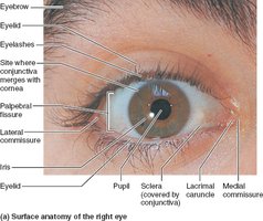
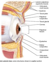
Accessory structures protect the eye and aid its function. These include the eyebrows, eyelids, conjunctiva, lacrimal apparatus, and extrinsic eye muscles.
Eyebrows: Shade the eyes from sunlight and prevent perspiration from reaching the eyes.
Eyelids (Palpebrae): Protect the eyes anteriorly, blink reflexively to moisten and protect the eye, and contain lubricating glands.
Conjunctiva: Transparent mucous membrane that lines the eyelids and covers the white of the eye, producing lubricating mucus.
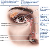
Lacrimal Apparatus: Produces and drains tears, which contain mucus, antibodies, and lysozyme for eye protection.
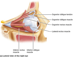
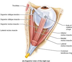
Extrinsic Eye Muscles: Six muscles that move the eyeball and help maintain its shape.
Extrinsic Eye Muscles
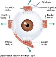
These muscles originate from the bony orbit and insert on the eyeball, allowing precise movement. Four rectus muscles (superior, inferior, lateral, medial) and two oblique muscles (superior, inferior) control eye movement.
Muscle | Action | Controlling Cranial Nerve |
|---|---|---|
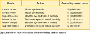
Lateral rectus | Moves eye laterally | VI (abducens) |
Medial rectus | Moves eye medially | III (oculomotor) |
Superior rectus | Elevates eye and turns it medially | III (oculomotor) |
Inferior rectus | Depresses eye and turns it medially | III (oculomotor) |
Inferior oblique | Elevates eye and turns it laterally | III (oculomotor) |
Superior oblique | Depresses eye and turns it laterally | IV (trochlear) |
Structure of the Eyeball
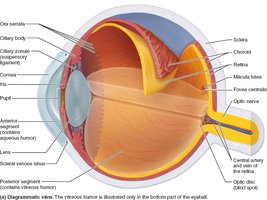
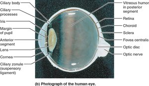
The eyeball consists of three layers (fibrous, vascular, inner), a lens, and internal fluids (humors). The lens divides the eye into anterior and posterior segments.
Fibrous Layer
Sclera: Opaque, white, posterior region; protects and shapes the eyeball.
Cornea: Transparent anterior portion; allows light entry and bends light.
Vascular Layer (Uvea)
Choroid: Pigmented, vascular layer; supplies blood and absorbs light.
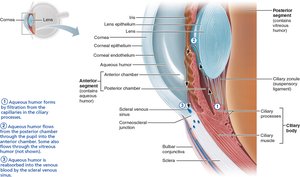
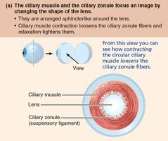
Ciliary Body: Contains ciliary muscles (control lens shape), ciliary processes (secrete aqueous humor), and ciliary zonule (holds lens).
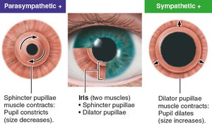
Iris: Colored part; controls pupil size via sphincter and dilator muscles.
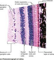
Inner Layer (Retina)
Pigmented Layer: Absorbs light, stores vitamin A, and phagocytizes debris.
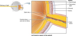
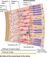
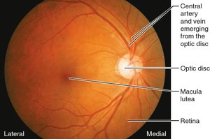
Neural Layer: Contains photoreceptors (rods and cones), bipolar cells, and ganglion cells. The optic disc is the blind spot where the optic nerve exits.
Photoreceptors
Rods: Sensitive to dim light; provide peripheral and night vision; no color discrimination.
Cones: Operate in bright light; provide high-resolution color vision; concentrated in the fovea centralis.
Blood Supply
Choroid supplies the outer third of the retina.
Central artery and vein supply the inner two-thirds.
Internal Chambers and Fluids
The lens and ciliary zonule divide the eye into anterior and posterior segments. The posterior segment contains vitreous humor, while the anterior segment contains aqueous humor, which is continuously formed and drained.
Lens
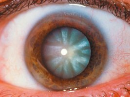
The lens is a biconvex, transparent, flexible structure that focuses light on the retina. With age, the lens becomes denser and less elastic, leading to presbyopia and increased risk of cataracts.
Light and Optics
Wavelength and Color
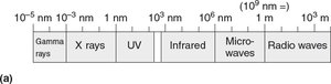
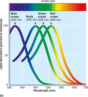
Visible light is a small part of the electromagnetic spectrum (400–700 nm). The color perceived depends on the wavelength reflected by objects.
Refraction and Lenses
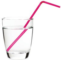
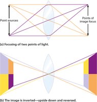
Refraction is the bending of light as it passes through different media. The cornea and lens refract light to focus it on the retina. Convex lenses converge light rays, while concave lenses diverge them.
Focusing Light on the Retina
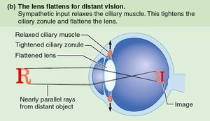
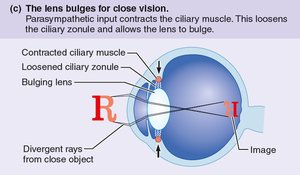
Light is refracted three times: entering the cornea, entering the lens, and leaving the lens. The cornea provides most of the refractive power, but the lens fine-tunes focus for distant and close vision.
Common Refraction Problems
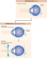
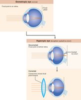
Myopia (Nearsightedness): Eyeball too long; focal point in front of retina; corrected with concave lenses.
Hyperopia (Farsightedness): Eyeball too short; focal point behind retina; corrected with convex lenses.
Astigmatism: Unequal curvature of cornea or lens; corrected with cylindrical lenses or laser procedures.
Phototransduction
Photoreceptor Structure and Function
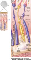
Photoreceptors (rods and cones) convert light into electrical signals. Each has an outer segment (containing visual pigments) and an inner segment (containing the cell body and synaptic terminal).
Rod vs. Cone Vision
Rods: High sensitivity, low acuity, monochromatic vision, many rods converge on one ganglion cell.
Cones: Low sensitivity, high acuity, color vision, one or few cones per ganglion cell.
Visual Pigments and Phototransduction
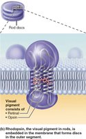
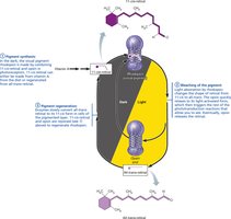
Visual pigments consist of retinal (from vitamin A) and opsins. Rods contain rhodopsin; cones contain one of three opsins (red, green, blue). Light absorption causes retinal to change shape, triggering a cascade that leads to hyperpolarization of the photoreceptor and signal transmission to the brain.
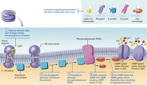
Signal Transmission in the Retina
Light hyperpolarizes photoreceptors, reducing glutamate release. This disinhibits bipolar cells, which then depolarize and stimulate ganglion cells to fire action potentials to the brain.
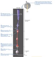
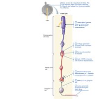
Visual Pathways and Processing
Visual Pathway to the Brain
Axons of ganglion cells form the optic nerve, which partially crosses at the optic chiasma. Visual information is relayed to the thalamus and then to the primary visual cortex for conscious perception. Some fibers project to midbrain regions for reflexes and circadian rhythms.
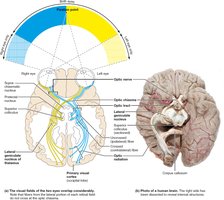
Smell (Olfaction)
Olfactory Epithelium and Receptors
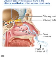
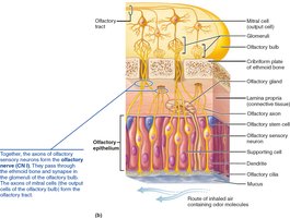
The olfactory epithelium in the nasal cavity contains bipolar olfactory sensory neurons with cilia that detect odorants dissolved in mucus. Olfactory neurons are unique in their ability to regenerate throughout life.
Physiology of Smell
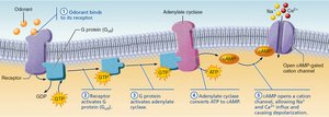
Odorants must be volatile and dissolve in mucus to bind to receptors, activating a G protein pathway that leads to depolarization and action potentials. Olfactory adaptation occurs with prolonged exposure to an odor.
Taste (Gustation)
Taste Buds and Papillae
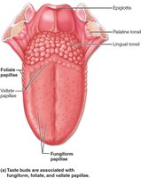
Taste buds are sensory organs located mainly on the tongue's papillae (fungiform, foliate, vallate). Each taste bud contains gustatory epithelial cells (taste receptors) and basal cells (stem cells).
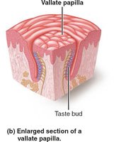
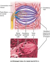
Basic Taste Sensations
Sweet: Sugars, alcohols, some amino acids
Sour: Acids (hydrogen ions)
Salty: Metal ions (e.g., Na+)
Bitter: Alkaloids (e.g., quinine, nicotine)
Umami: Amino acids (glutamate, aspartate)
There is evidence for a sixth taste for long-chain fatty acids.
Physiology of Taste
Tastants must dissolve in saliva, diffuse into taste pores, and contact gustatory hairs. Different tastes activate different transduction mechanisms, often involving ion channels or G protein-coupled receptors (gustducin).
Hearing and Equilibrium: The Ear
Structure of the Ear
The ear is divided into three regions: external (auricle and auditory canal), middle (tympanic cavity and ossicles), and internal (bony and membranous labyrinths). The internal ear contains the cochlea (hearing) and vestibular apparatus (equilibrium).
Sound Transmission and Transduction
Sound waves cause the tympanic membrane to vibrate, which is transmitted via ossicles to the oval window, creating fluid waves in the cochlea. Hair cells in the cochlear duct transduce these waves into electrical signals sent to the brain.
Equilibrium
Equilibrium receptors in the vestibule (maculae) and semicircular canals (cristae ampullares) detect linear and rotational movements, respectively, helping maintain balance and posture.
Clinical Applications and Homeostatic Imbalances
Chalazion: Obstructed tarsal gland; painless bump.
Sty: Inflammation of sebaceous gland at eyelash base; painful.
Conjunctivitis/Pinkeye: Inflammation/infection of conjunctiva; highly contagious.
Glaucoma: Increased intraocular pressure; can lead to blindness.
Cataract: Clouding of the lens; impairs vision.
Myopia/Hyperopia/Astigmatism: Refraction disorders; corrected with lenses or surgery.
Color Blindness: Inherited lack of cone pigments; more common in males.
Nyctalopia: Night blindness due to rod degeneration, often from vitamin A deficiency.
Anosmias: Olfactory disorders from injury, infection, or neurological disease.
Taste Disorders: Less common; may result from infection, injury, or medications.
Additional info: This summary covers the anatomy and physiology of the special senses, focusing on the structure and function of the eye, ear, and associated sensory pathways, as well as clinical correlations relevant to these systems.
