 뒤로
뒤로Chapter 17: Blood – Structure, Function, and Disorders
스터디 가이드 - 스마트 노트
 자료에 맞춘 맞춤형 노트, 핵심 정의, 예시, 맥락을 확장해 제공합니다.
Blood: Overview and Functions
Functions of Blood
Blood is a vital connective tissue that performs several essential functions in the human body:
Transport: Blood transports oxygen (O2), carbon dioxide (CO2), nutrients, metabolic wastes, hormones, and heat throughout the body.
Protection: White blood cells (WBCs), antibodies, and platelets in blood help defend the body against infection and initiate clotting to prevent blood loss.
Regulation: Blood regulates fluid balance, pH buffering, and body temperature.
Properties of Blood
Physical and Chemical Properties
Temperature: 38°C (100.4°F), slightly higher than body temperature due to friction and metabolic activity.
pH: Maintained between 7.35 and 7.45, making blood slightly alkaline.
Osmolarity: The total molar concentration of dissolved particles (mainly sodium ions, proteins, and RBCs). High osmolarity draws fluid into the blood, raising blood pressure; low osmolarity causes fluid retention in tissues, leading to edema and decreased blood pressure.
Volume: 5–6 liters in males, 4–5 liters in females.
Viscosity: Blood is about five times as viscous as water, which affects its flow through vessels.
Oncotic Pressure
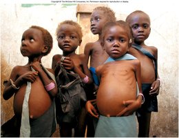
Oncotic pressure is a form of osmotic pressure exerted by plasma proteins, primarily albumin, which pulls water into the circulatory system. When oncotic pressure is pathologically low (e.g., in kwashiorkor due to starvation), blood pressure drops and edema develops.
Hematopoiesis: Blood Cell Formation
Embryonic and Fetal Hematopoiesis
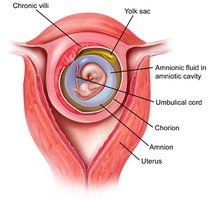
During embryonic and fetal development, blood cell formation (hematopoiesis) begins in the yolk sac, which produces stem cells that colonize the liver, bone marrow, spleen, and thymus.
Adult Hematopoiesis
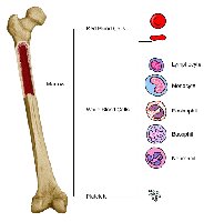
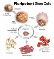
After birth, hematopoiesis occurs primarily in the red bone marrow, which contains pluripotent stem cells capable of giving rise to all blood cell types.
Stem Cell Differentiation
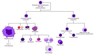
Pluripotent stem cells differentiate into two main lines:
Myeloid line: Produces erythrocytes (RBCs), platelets, and most leukocytes (granulocytes and monocytes).
Lymphoid line: Produces lymphocytes (B cells, T cells, and natural killer cells).
Blood Components
Hematocrit
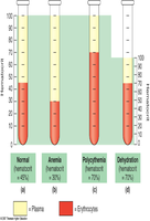
Hematocrit is the percentage of blood volume occupied by erythrocytes (RBCs). It is a key indicator of oxygen-carrying capacity and varies with health, hydration, and disease states.
Plasma Composition
Plasma is the liquid portion of blood, consisting of water, proteins, nutrients, gases, and wastes. Serum is plasma without fibrinogen (a clotting protein).
Albumins: Most abundant plasma protein; contributes to viscosity and osmolarity.
Globulins: Antibodies that provide immune functions.
Fibrinogen: Precursor to fibrin, essential for blood clotting.
Nitrogenous compounds: Includes amino acids and nitrogenous wastes (e.g., urea).
Nutrients: Glucose, vitamins, fats, minerals.
Gases: O2 and CO2.
Electrolytes: Sodium, potassium, calcium, etc.
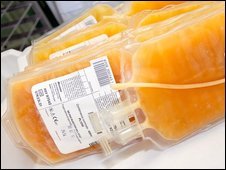
Formed Elements
The formed elements of blood include erythrocytes (RBCs), leukocytes (WBCs), and platelets.
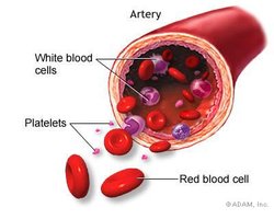
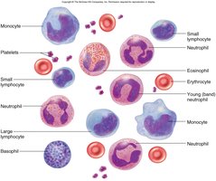
Erythrocytes (Red Blood Cells)
Structure and Function
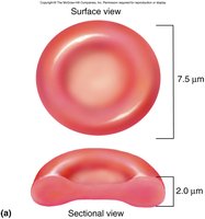
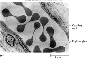
Erythrocytes are disc-shaped, anucleate cells with a biconcave shape that increases surface area for gas exchange. They transport oxygen and some carbon dioxide and have a lifespan of about 120 days.
Hemoglobin Structure and Function
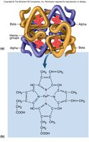
Hemoglobin (Hb) is the main protein in RBCs, responsible for oxygen transport. Each molecule consists of four globin chains (2 alpha, 2 beta) and four heme groups, each binding one O2 molecule. Hemoglobin also transports some CO2.
1 hemoglobin molecule can carry 4 O2 molecules.
About 33% of RBC cytoplasm is hemoglobin.
Gender Differences in RBC Parameters
Parameter | Males | Females |
|---|---|---|
Hematocrit (%) | 42–52 | 37–48 |
Hemoglobin (g/dL) | 13–18 | 12–16 |
RBC count (million/μL) | 4.6–6.2 | 4.2–5.4 |
Additional info: Differences are due to androgens (which stimulate RBC production), menstrual losses, and body fat percentage.
Erythropoiesis: RBC Production
Hormonal Regulation
Erythropoietin (EPO), a hormone produced by the kidneys, stimulates the formation of RBCs in response to low oxygen levels (hypoxia), increased exercise, or high altitude.


Negative Feedback Control
A drop in RBC count triggers the kidneys to secrete EPO, which stimulates bone marrow to increase RBC production. The process takes 3–4 days for RBC count to rise.
Stages of Erythrocyte Production
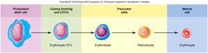
Development of RBCs takes 3–5 days and involves several stages:
Pluripotent stem cell
Erythrocyte colony-forming unit (CFU) – first committed cell, has EPO receptors
Erythroblast – multiplies and synthesizes hemoglobin
Reticulocyte – nucleus is discarded
Mature erythrocyte
Nutritional Requirements for Erythropoiesis
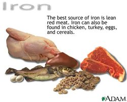
Iron: Essential for hemoglobin synthesis; stomach acid converts Fe3+ to absorbable Fe2+.
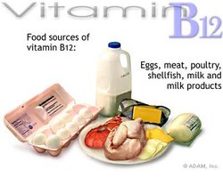
Vitamin B12: Absorbed with the help of intrinsic factor from the stomach; necessary for DNA synthesis in RBCs.
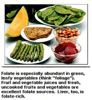
Folate (Folic acid): Required for cell division and maturation of RBCs.
RBC Life Cycle and Disorders
RBC Life Cycle
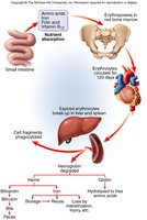
RBCs circulate for about 120 days before being broken down in the liver and spleen. Hemoglobin is degraded, and its components are recycled or excreted.
Polycythemia
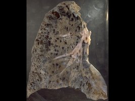
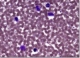
Polycythemia is an excess of RBCs. It can be primary (due to cancer of erythropoietic cell line) or secondary (due to dehydration, emphysema, high altitude, or physical conditioning). Dangers include increased blood volume, pressure, and viscosity, which can lead to embolism, stroke, or heart failure.
Anemia
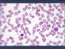
Anemia is a deficiency of RBCs or hemoglobin. Causes include:
Inadequate erythropoiesis or hemoglobin synthesis (e.g., vitamin B12 or iron deficiency, kidney failure, aplastic anemia)
Blood loss or destruction
Effects include tissue hypoxia, low blood osmolarity (leading to edema), and low blood viscosity (causing increased heart rate and decreased blood pressure).
Sickle-Cell Disease
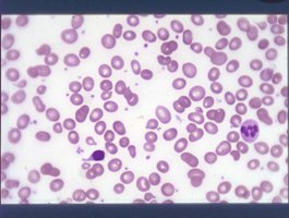
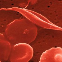
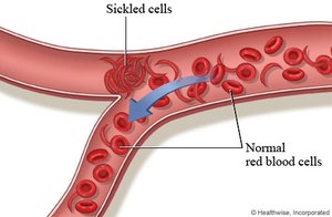
Sickle-cell disease is a hereditary disorder caused by a single amino acid substitution in the hemoglobin beta chain, resulting in hemoglobin S (HbS). In low oxygen conditions, HbS polymerizes, causing RBCs to become sickle-shaped, sticky, and prone to blocking vessels. This leads to pain, organ damage, and reduced lifespan.
Sickle-cell trait (heterozygous for HbS) confers resistance to malaria, explaining its persistence in populations where malaria is endemic.
