 뒤로
뒤로Chapter 17: The Special Senses – Study Notes for Anatomy & Physiology
스터디 가이드 - 스마트 노트
 자료에 맞춘 맞춤형 노트, 핵심 정의, 예시, 맥락을 확장해 제공합니다.
An Introduction to the Special Senses
The special senses include olfaction (smell), gustation (taste), vision, equilibrium (balance), and hearing. These senses are mediated by specialized organs and pathways that allow the body to detect and interpret complex environmental stimuli.
Olfaction (The Sense of Smell)
Olfactory Organs and Structure
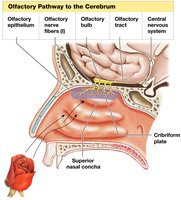
The olfactory organs are located in the nasal cavity and consist of two main layers: the olfactory epithelium and the lamina propria. Odorants are small, airborne, organic molecules that stimulate olfactory receptors.
Olfactory epithelium: Covers the inferior surface of the cribriform plate, superior portion of the perpendicular plate, and superior nasal conchae of the ethmoid bone.
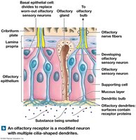
Lamina propria: Underlies the olfactory epithelium; contains areolar tissue, blood vessels, nerves, and olfactory glands that secrete mucus.
Olfactory Sensory Neurons
Olfactory sensory neurons: Highly modified nerve cells with dendrites extending into the mucus layer; their axons form the olfactory nerves.
Supporting cells and basal epithelial cells (stem cells) are also present.
Physiology of Olfaction
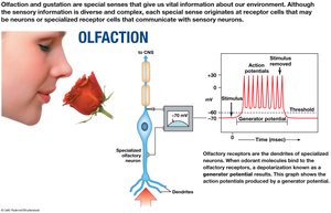
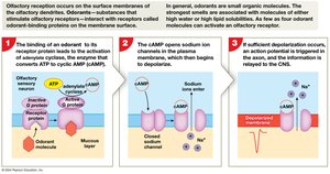
Olfaction begins when an odorant binds to a G protein–coupled receptor on the olfactory dendrite, activating a second messenger (cAMP) that opens sodium ion channels, leading to depolarization and action potential generation.
Olfactory Pathways and Discrimination
Axons of olfactory sensory neurons penetrate the cribriform plate and synapse in the olfactory bulbs.
Olfactory bulb neurons form the olfactory tracts, which project to the olfactory cortex, hypothalamus, and limbic system.
Olfactory information reaches the cerebral cortex without passing through the thalamus.
Humans can distinguish 2,000–4,000 odorants; olfactory neurons are replaced frequently, but their number declines with age.
Gustation (The Sense of Taste)
Taste Buds and Lingual Papillae
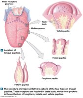
Gustation provides information about foods and liquids. Taste receptor cells are found in taste buds, which are located on the superior surface of the tongue and parts of the pharynx and larynx. Taste buds are associated with different types of lingual papillae:
Filiform papillae: Provide friction, do not contain taste buds.
Fungiform papillae: Contain about five taste buds each.
Vallate papillae: Contain about 100 taste buds each, arranged in a V-shape at the posterior tongue.
Foliate papillae: Found on the lateral margins, contain taste buds.
Taste Bud Structure
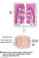
Basal epithelial cells: Stem cells for taste bud regeneration.
Transitional cells: Supporting cells.
Taste receptor cells: Extend microvilli (taste hairs) through a taste pore and synapse with sensory neurons.
Gustatory Pathways
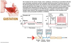
Taste receptor cells release neurotransmitters that stimulate sensory neurons of the facial, glossopharyngeal, and vagus nerves.
Sensory fibers synapse in the solitary nucleus of the medulla oblongata, then ascend to the thalamus and gustatory cortex in the insula.
Taste perception is influenced by texture and olfactory information.
Gustatory Discrimination
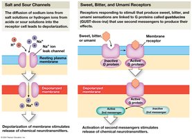
Primary taste sensations: Sweet, salty, sour, bitter.
Additional sensations: Umami (savory, glutamate), water (detected in the pharynx).
Different tastes involve different receptor mechanisms: salty and sour via ion channels; sweet, bitter, and umami via G protein–coupled receptors.
Taste sensitivity varies among individuals and is often inherited. Children have more taste receptors than adults, and the number declines with age.
Structures of the Eye
Accessory Structures
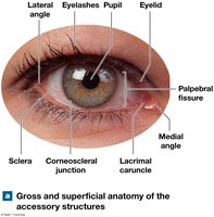
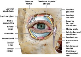
The eye is protected and supported by accessory structures, including the eyelids, superficial epithelium, and lacrimal apparatus.
Eyelids (palpebrae): Protect and lubricate the eye; contain tarsal glands and eyelashes.
Conjunctiva: Epithelial membrane covering the inner eyelids and anterior eye surface.
Lacrimal apparatus: Produces, distributes, and removes tears.
Anatomy of the Eyeball
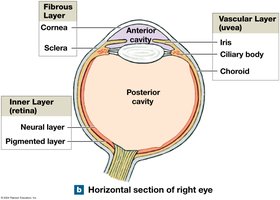
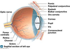
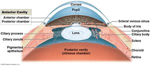
Fibrous layer: Sclera (white of the eye) and cornea (transparent anterior part).
Vascular layer (uvea): Iris (controls pupil size), ciliary body (controls lens shape), and choroid (provides nutrients to retina).
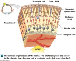
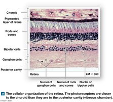
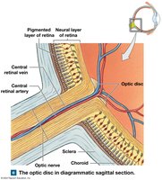
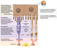
Inner layer (retina): Pigmented layer (absorbs light) and neural layer (contains photoreceptors).
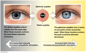
Pupillary Muscles
Sphincter pupillae: Constricts pupil (parasympathetic control).
Dilator pupillae: Dilates pupil (sympathetic control).
Retina and Photoreceptors
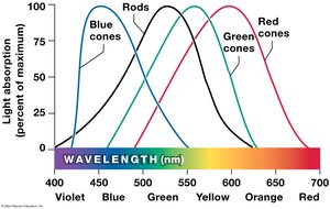
Rods: Sensitive to low light, do not detect color, more numerous in peripheral retina.
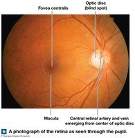
Cones: Detect color and provide sharp vision, concentrated in the macula and fovea centralis.
Bipolar cells: Synapse with rods and cones.
Ganglion cells: Axons form the optic nerve.
Optic Disc and Clinical Correlates
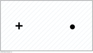
Optic disc: Origin of the optic nerve, lacks photoreceptors (blind spot).
Diseases: Diabetic retinopathy, detached retina.
Chambers and Fluids of the Eye
Aqueous humor: Circulates in the anterior cavity, provides nutrients, maintains intraocular pressure.
Vitreous body: Gelatinous mass in the posterior cavity, stabilizes eye shape.
Lens: Focuses visual image on photoreceptors; loss of transparency leads to cataracts.
Formation of a Visual Image
Refraction and Focusing
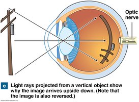
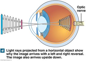
Light is refracted by the cornea and lens to focus images on the retina. The lens changes shape (accommodation) to focus on near or distant objects.
Focal distance: Distance from the lens to the focal point; affected by object distance and lens shape.
Astigmatism: Irregular refraction causes distorted images.
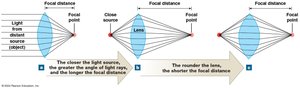
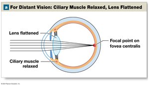
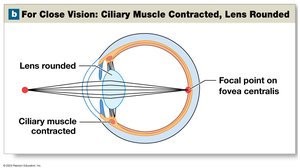
Image Formation and Visual Acuity
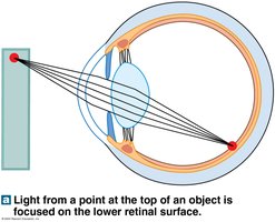
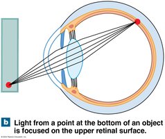
The image on the retina is inverted and reversed; the brain corrects this orientation.
Visual acuity: Clarity of vision; legal blindness is defined as visual acuity less than 20/200.
Scotoma: Permanent blind spot; floaters: Debris in the vitreous body.
Physiology of Vision
Photoreceptors and Visual Pigments
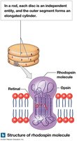
Rods: Contain rhodopsin (opsin + retinal, derived from vitamin A).
Cones: Contain different opsins sensitive to blue, green, or red wavelengths.
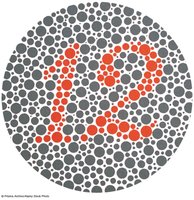
Color blindness: Inability to distinguish certain colors due to missing cones.
Photoreception and Signal Transduction
In darkness, rods are depolarized due to open sodium channels (maintained by cGMP) and release glutamate.
Light absorption changes retinal from 11-cis to 11-trans form, activating transducin and phosphodiesterase, which closes sodium channels, hyperpolarizing the cell.
Bleaching: Retinal and opsin dissociate after photon absorption; retinal is converted back to 11-cis form and recombines with opsin.
Vitamin A deficiency can cause night blindness.
Light and Dark Adaptation
Dark-adapted state: Visual pigments are fully receptive.
Light-adapted state: Bleaching and reassembly of pigments are balanced.
Pupil size adjusts to control light entry.
Visual Pathways and Processing
Photoreceptors → bipolar cells → ganglion cells (optic nerve).
Optic nerves partially cross at the optic chiasm, then project to the lateral geniculate bodies and visual cortex.
M cells: Monitor rods, detect motion and general form.
P cells: Monitor cones, detect fine detail and color.
Depth perception arises from comparing images from both eyes.
The Ear: Hearing and Equilibrium
Anatomy of the Ear
External ear: Auricle (pinna), external acoustic meatus, tympanic membrane.
Middle ear: Auditory ossicles (malleus, incus, stapes), auditory tube, muscles (tensor tympani, stapedius).
Internal ear: Bony and membranous labyrinths, vestibule, semicircular canals, cochlea.
Equilibrium
Detected by hair cells in the vestibular complex (vestibule and semicircular canals).
Semicircular ducts detect rotational movement; utricle and saccule detect linear acceleration and gravity.
Otoliths in the otolithic membrane enhance sensitivity to gravity and acceleration.
Hearing
Sound waves vibrate the tympanic membrane, which moves the ossicles and creates pressure waves in the cochlear fluids.
Hair cells in the spiral organ (organ of Corti) detect these vibrations and convert them to nerve impulses.
Pitch is determined by the location of basilar membrane vibration; loudness by the amplitude of vibration.
Auditory Pathways
Sensory neurons in the spiral ganglion form the cochlear nerve, which projects to the cochlear nucleus, superior olivary nuclei, inferior colliculi, medial geniculate body, and finally the auditory cortex.
Special Sense | Receptor Type | Main Organ | Main Pathway |
|---|---|---|---|
Olfaction | Olfactory neurons | Nasal cavity | Olfactory bulb → tract → cortex |
Gustation | Taste receptor cells | Taste buds (tongue) | Cranial nerves → medulla → thalamus → cortex |
Vision | Rods & cones | Retina (eye) | Optic nerve → chiasm → thalamus → cortex |
Equilibrium | Hair cells | Vestibule, semicircular canals | Vestibular nerve → brainstem → cerebellum/cortex |
Hearing | Hair cells | Cochlea | Cochlear nerve → brainstem → cortex |
Additional info: This summary integrates and expands upon the provided lecture slides, ensuring all major concepts and structures are explained with academic clarity and completeness for college-level Anatomy & Physiology students.
