 뒤로
뒤로Chapter 4: Histology – The Study of Tissues
스터디 가이드 - 스마트 노트
 자료에 맞춘 맞춤형 노트, 핵심 정의, 예시, 맥락을 확장해 제공합니다.
Histology: The Study of Tissues
Introduction to Tissues
Histology is the study of tissues, which are groups of structurally and functionally related cells and their external environment working together to perform common functions. All tissues share two basic components: a discrete population of cells and the surrounding material known as the extracellular matrix (ECM).
Tissue: Group of related cells and their ECM performing a common function.
Histology: Study of normal tissue structure.
Extracellular Matrix (ECM): Provides strength, directs cell placement, regulates cell activity, and holds cells in position.
Types of Tissues
Epithelial Tissues: Sheets of tightly packed cells with little ECM; cover and line surfaces and form glands.
Connective Tissues: Bind, support, protect, and allow transport; cells are scattered in ECM.
Muscle Tissues: Cells contract to generate force; little ECM.
Nervous Tissues: Neurons generate, send, and receive messages; supported by neuroglial cells and unique ECM.
Extracellular Matrix (ECM)
Components of the ECM
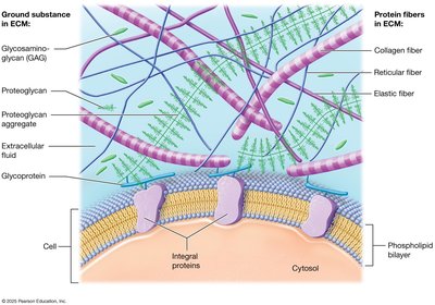
The ECM is composed of ground substance and protein fibers, each contributing to tissue structure and function.
Ground Substance: Gel-like material with extracellular fluid, water, ions, nutrients, and macromolecules.
Protein Fibers: Provide tensile strength and support.
Ground Substance Macromolecules
Glycosaminoglycans (GAGs): Negatively charged polysaccharides (e.g., chondroitin sulfate, hyaluronic acid) that attract water and ions.
Proteoglycans: GAGs attached to protein cores; aggregate to resist compression and act as diffusion barriers.
Glycoproteins (Cell-Adhesion Molecules, CAMs): Bind cell surface proteins and fibers, maintaining tissue architecture.
Protein Fibers in ECM
Collagen Fibers: Strong, resistant to tension and pressure; most abundant protein in the body.
Elastic Fibers: Made of elastin; allow stretch and recoil (distensibility and elasticity).
Reticular Fibers: Thin collagen fibers forming supportive networks in tissues and organs.
Cell Junctions
Types of Cell Junctions
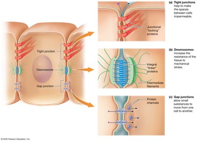
Cell junctions are connections between neighboring cells, crucial for tissue integrity and communication.
Tight (Occluding) Junctions: Integral proteins create a seal, preventing passage of macromolecules.
Desmosomes: Button-like junctions distributing mechanical stress via linker proteins and intermediate filaments.
Gap Junctions: Protein channels allowing small substances to pass between cells for communication.
Epithelial Tissues
Functions of Epithelial Tissue
Protection: Shields underlying tissues; produces keratin; rapid mitosis.
Immune Defense: Contains immune cells.
Secretion: Forms glands producing sweat, oil, hormones.
Transport: Selectively permeable barriers for substance movement.
Sensation: Supplied with nerves; specialized cells detect stimuli.
Components and Classification of Epithelia
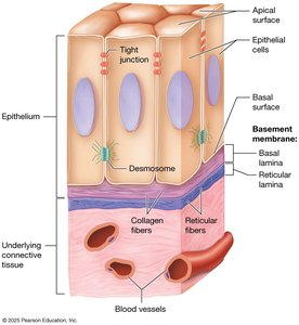
Basement Membrane: Anchors epithelium to connective tissue; consists of basal lamina (epithelial ECM) and reticular lamina (connective tissue ECM).
Cell Surfaces: Apical (free), basal (attached), and lateral (side) surfaces.
Classification by Layers and Shape
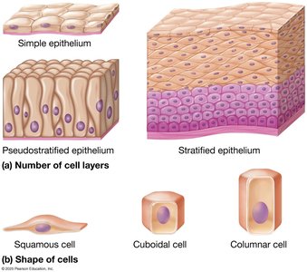
Number of Layers:
Simple: One layer
Stratified: Multiple layers
Pseudostratified: Appears multilayered but is a single layer
Cell Shape:
Squamous: Flattened
Cuboidal: Cube-shaped
Columnar: Tall and elongated
Covering and Lining Epithelia
Simple Epithelia
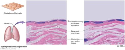
Simple Squamous: Single layer of flat cells; rapid diffusion; found in lungs, serous membranes, blood vessels.
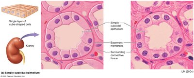
Simple Cuboidal: Single layer of cube-shaped cells; secretion and absorption; found in kidney tubules, glands.
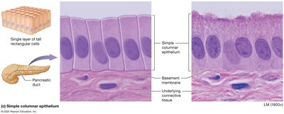
Simple Columnar: Single layer of tall cells; absorption, secretion, may have microvilli or cilia; found in intestines, uterine tubes.
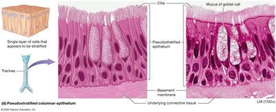
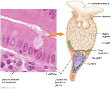
Pseudostratified Columnar: Appears stratified; nuclei at different heights; often ciliated with goblet cells; found in respiratory passages.
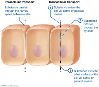
Transport Across Simple Epithelia
Paracellular Transport: Substances pass between cells.
Transcellular Transport: Substances move through cells (enter, diffuse, exit).
Stratified Epithelia
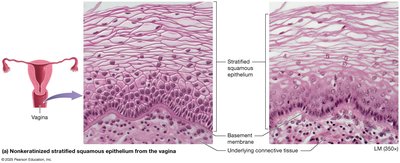
Stratified Squamous (Nonkeratinized): Multiple layers; protection; found in mouth, esophagus, vagina.
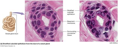
Stratified Cuboidal: Two layers of cuboidal cells; rare; lines sweat gland ducts.
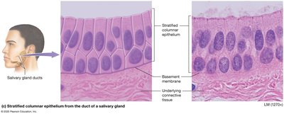
Stratified Columnar: Few layers; columnar apical, cuboidal basal; rare; found in salivary gland ducts, male urethra.
Transitional Epithelium: Basal cells cuboidal, apical cells dome-shaped (relaxed) or squamous (stretched); found in urinary tract.
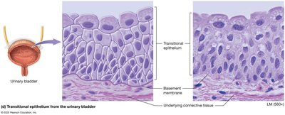
Summary Tables of Epithelial Tissues
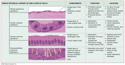
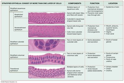
Glandular Epithelia
Types of Glands
Exocrine Glands: Secrete products via ducts to epithelial surfaces; local action.
Endocrine Glands: Secrete hormones directly into blood; act on distant targets.
Exocrine Gland Structure
Unicellular: Goblet cells secrete mucus in digestive and respiratory tracts.
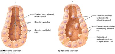
Modes of Secretion
Merocrine: Products released by exocytosis (e.g., sweat, salivary glands).
Holocrine: Products released by cell rupture (e.g., sebaceous glands).
Apocrine: Products released with part of cytoplasm (e.g., mammary glands).
Concept Boost: Histological Identification
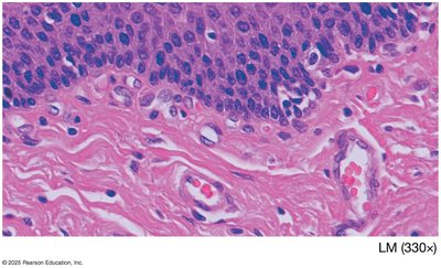
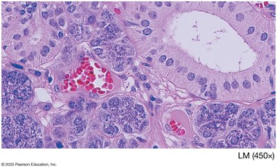
Microscopic Appearance of Tissues
Histological sections are stained, often appearing pink due to dyes.
Cell nuclei stain dark purple; ECM appears clear or lightly colored; protein fibers are wavy or straight lines.
Carcinomas and Epithelial Cancer
Carcinogens and Carcinomas
Carcinogens: Agents causing DNA changes that may lead to cancer.
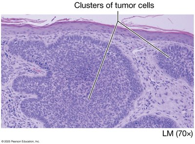
Carcinomas: Cancers of epithelial tissue; e.g., basal cell carcinoma of the skin.
Basement membrane: Can slow cancer spread; if not penetrated, called carcinoma in situ.
Additional info:
Further details on connective, muscle, and nervous tissues, as well as tissue repair and membranes, are covered in subsequent sections of the chapter.
