 뒤로
뒤로Chapter 4: The Tissue Level of Organization – Study Notes
스터디 가이드 - 스마트 노트
 자료에 맞춘 맞춤형 노트, 핵심 정의, 예시, 맥락을 확장해 제공합니다.
The Tissue Level of Organization
An Introduction to Tissues
Tissues are collections of specialized cells and cell products that perform specific functions. They possess distinct structural and functional properties and, when combined, form organs such as the heart or liver. The study of tissues is known as histology.
Tissues are the building blocks of organs and organ systems.
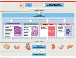
There are four primary tissue types: epithelial, connective, muscle, and nervous tissue.
Types of Tissue
The Four Basic Tissue Types
Epithelial tissue: Covers exposed surfaces, lines internal passageways, and forms glands.
Connective tissue: Fills internal spaces, supports other tissues, transports materials, and stores energy.
Muscle tissue: Specialized for contraction, producing movement.
Nervous tissue: Carries electrical signals from one part of the body to another.
Epithelial Tissue
Characteristics and Functions
Epithelial tissue includes layers of cells that cover internal or external surfaces and form glands. It serves several essential functions:
Physical protection: Protects against abrasion, dehydration, and destruction by chemical or biological agents.
Control permeability: Regulates the movement of substances into and out of the body.
Provide sensation: Contains sensory nerve endings for detecting stimuli.
Produce specialized secretions: Glandular epithelium releases secretions onto surfaces or into the blood.
Structural Characteristics
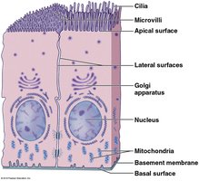
Polarity: Has apical (exposed) and basal (attached) surfaces.
Cellularity: Composed almost entirely of tightly packed cells connected by cell junctions.
Attachment: Basal surface attached to a basement membrane.
Avascularity: Lacks blood vessels; nutrients diffuse from underlying tissues.
Regeneration: High capacity for cell division and repair.
Specializations of Epithelial Cells
Move fluids over the epithelium (protection)
Move fluids through the epithelium (permeability)
Produce secretions (protection and messaging)
Apical surface may have microvilli (increase absorption/secretion) or cilia (move fluids)
Integrity of Epithelia
The integrity of epithelial tissue is maintained by intercellular connections, attachment to the basement membrane, and ongoing maintenance and repair.
Cell adhesion molecules (CAMs): Transmembrane proteins that connect cells to each other and to the basement membrane.
Proteoglycans: Act as intercellular cement.
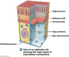
Cell junctions: Specialized structures that connect cells (gap junctions, tight junctions, desmosomes).
Types of Cell Junctions
Gap junctions: Allow rapid communication and passage of ions/small molecules between cells.
Tight junctions: Prevent passage of water and solutes between cells; maintain distinct environments.
Desmosomes: Provide strong attachment between cells; resist mechanical stress.
Hemidesmosomes: Attach cells to the basement membrane.
Classification of Epithelia
By Shape and Layers
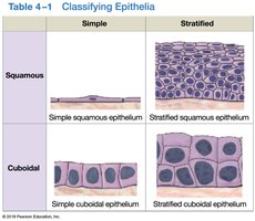
Shape: Squamous (thin, flat), Cuboidal (cube-shaped), Columnar (tall, rectangular)
Layers: Simple (single layer), Stratified (multiple layers)
Simple | Stratified | |
|---|---|---|
Squamous | Simple squamous epithelium | Stratified squamous epithelium |
Cuboidal | Simple cuboidal epithelium | Stratified cuboidal epithelium |
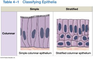
Columnar | Simple columnar epithelium | Stratified columnar epithelium |
Squamous Epithelia
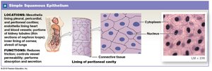
Simple squamous: Absorption and diffusion; lines body cavities (mesothelium), heart, and blood vessels (endothelium).
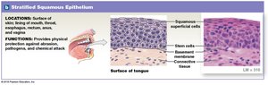
Stratified squamous: Protects against mechanical stress; may be keratinized (skin) or non-keratinized (oral cavity, esophagus).
Cuboidal Epithelia
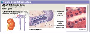
Simple cuboidal: Secretion and absorption; found in glands, kidney tubules.
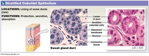
Stratified cuboidal: Rare; found in ducts of sweat and mammary glands.
Transitional Epithelium
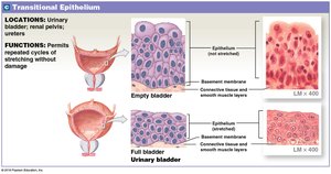
Transitional epithelium tolerates repeated cycles of stretching and recoiling without damage. It is found in the urinary bladder.
Columnar Epithelia
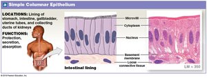
Simple columnar: Absorption and secretion; found in stomach, intestines.
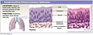
Pseudostratified columnar: Appears layered but all cells contact the basement membrane; often ciliated; found in respiratory tract.
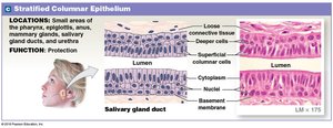
Stratified columnar: Rare; provides protection in pharynx, anus, urethra.
Glandular Epithelia
Endocrine glands: Release hormones into the bloodstream; ductless.
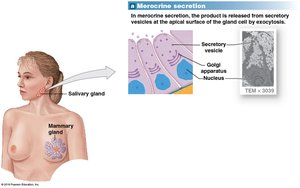
Exocrine glands: Release secretions onto epithelial surfaces through ducts.
Methods of Secretion
Method | Description | Example |
|---|---|---|
Merocrine | Product released by exocytosis | Sweat glands, mucin |
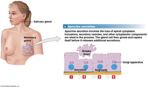
Apocrine | Loss of apical cytoplasm | Mammary glands |
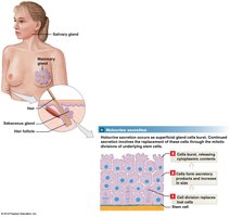
Holocrine | Cell bursts, releasing contents | Sebaceous glands |
Types of Exocrine Secretions
Serous glands: Watery secretions with enzymes (e.g., parotid salivary gland).
Mucous glands: Secrete mucins that hydrate to form mucus (e.g., submucosal glands of small intestine).
Mixed exocrine glands: Both serous and mucous (e.g., submandibular salivary gland).
Connective Tissue
Components and Functions
Connective tissues consist of specialized cells, extracellular protein fibers, and ground substance. The matrix (fibers + ground substance) determines the tissue's function and volume.
Establish structural framework
Transport fluids and dissolved materials
Protect delicate organs
Support, surround, and interconnect other tissues
Store energy reserves (triglycerides)
Defend against microorganisms
Categories of Connective Tissue
Connective tissue proper: Connects and protects (e.g., adipose, tendons)
Fluid connective tissues: Transport (blood, lymph)
Supporting connective tissues: Structural strength (cartilage, bone)
Cells of Connective Tissue Proper
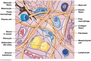
Fibroblasts: Secrete proteins and hyaluronan; most abundant.
Fibrocytes: Maintain connective tissue fibers.
Adipocytes: Store fat.
Mesenchymal cells: Stem cells for repair.
Melanocytes: Synthesize melanin pigment.
Macrophages: Phagocytic; engulf pathogens and debris.
Mast cells: Stimulate inflammation; release histamine and heparin.
Lymphocytes: Immune response; may become plasma cells (antibodies).
Microphages: Phagocytic blood cells (neutrophils, eosinophils).
Fibers of Connective Tissue Proper
Collagen fibers: Strong, flexible, resist force in one direction (tendons, ligaments).
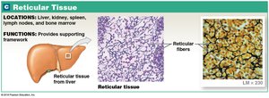
Reticular fibers: Network of interwoven fibers; support organs.
Elastic fibers: Contain elastin; stretch and return to original length (elastic ligaments).
Ground Substance
Clear, colorless, and viscous; fills spaces between cells and slows pathogen movement.
Embryonic Connective Tissues
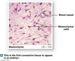
Mesenchyme: First connective tissue in embryos.
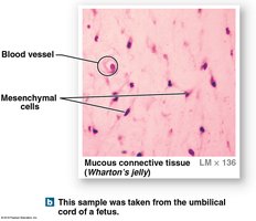
Mucous connective tissue: Loose tissue in umbilical cord (Wharton's jelly).
Loose Connective Tissues
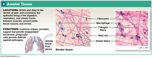
Areolar tissue: Cushions organs, provides support, allows movement.
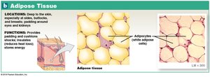
Adipose tissue: Stores fat, insulates, absorbs shocks.
Reticular tissue: Provides supporting framework for organs.
Dense Connective Tissues
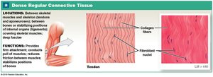
Dense regular: Parallel collagen fibers; tendons, ligaments.
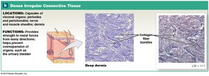
Dense irregular: Interwoven collagen fibers; dermis, organ capsules.
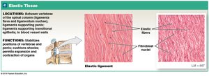
Elastic tissue: Elastic fibers; stabilizes vertebrae, cushions shocks.
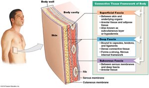
Fasciae
Connective tissue layers that support and surround organs. Three types: superficial, deep, and subserous fascia.
Fluid Connective Tissues
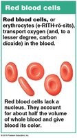
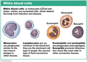
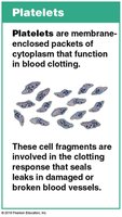
Blood: Watery matrix (plasma), formed elements (red cells, white cells, platelets).
Lymph: Interstitial fluid entering lymphatic vessels; immune surveillance.
Supporting Connective Tissues
Cartilage
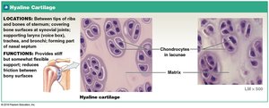
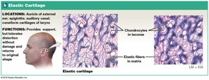
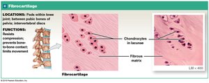
Matrix: Firm gel with chondroitin sulfates; cells are chondrocytes in lacunae.
Avascular: Receives nutrients by diffusion; perichondrium surrounds cartilage.
Types of Cartilage
Hyaline cartilage: Most common; reduces friction; found in joints, ribs, sternum, trachea.
Elastic cartilage: Flexible; found in external ear, epiglottis.
Fibrocartilage: Durable, resists compression; found in intervertebral discs, pubic symphysis.
Cartilage Growth
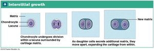
Interstitial growth: Growth from within the cartilage.
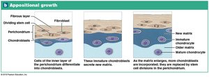
Appositional growth: Growth at the outer surface.
Bone (Osseous Tissue)
Strong, calcified matrix with collagen fibers; resists shattering.
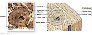
Osteocytes in lacunae, arranged around central canals.
Periosteum covers bone surfaces (fibrous and cellular layers).
Tissue Membranes
Types of Tissue Membranes
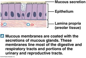
Mucous membranes: Line passageways with external connections; moist surfaces for absorption/secretion.
Serous membranes: Line cavities not open to the outside; secrete serous fluid to reduce friction.
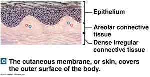
Cutaneous membrane: Skin; thick, waterproof, dry.
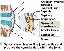
Synovial membranes: Line joint cavities; produce synovial fluid for lubrication.
Muscle Tissue
Types of Muscle Tissue
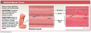
Skeletal muscle: Long, cylindrical, striated, multinucleate; voluntary movement.
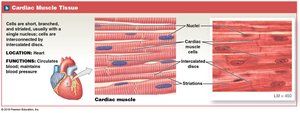
Cardiac muscle: Short, branched, striated; found only in heart; involuntary, connected by intercalated discs.
Smooth muscle: Small, spindle-shaped, non-striated; found in walls of hollow organs; involuntary.
Nervous Tissue
Structure and Function
Nervous tissue is specialized for conducting electrical impulses, rapidly sensing internal and external environments, and processing information. It is concentrated in the brain and spinal cord.
Neurons: Nerve cells that perform electrical communication.
Neuroglia: Supporting cells; repair and supply nutrients to neurons.
Parts of a Neuron
Cell body: Contains nucleus and nucleolus.
Dendrites: Receive incoming signals.
Axon: Carries outgoing electrical signals.
Tissue Injuries and Repair
Inflammation and Regeneration
Inflammation: First response to injury; signs include swelling, redness, heat, pain.
Regeneration: Replacement of damaged tissue; varies by tissue type (epithelia and connective tissues regenerate well; muscle and nervous tissue poorly).
Fibrosis: Replacement of damaged cardiac muscle cells by fibrous tissue (scar tissue).
Aging, Regeneration, and Cancer
Effects of Aging
Decreased speed and effectiveness of tissue repair.
Thinner epithelia, fragile connective tissues, increased bruising, brittle bones, cardiovascular disease, mental deterioration.
Increased cancer incidence with age; most cancers caused by chemical/environmental exposure.
