 뒤로
뒤로Chapter 4: Tissue – The Living Fabric (Anatomy & Physiology Study Notes)
스터디 가이드 - 스마트 노트
 자료에 맞춘 맞춤형 노트, 핵심 정의, 예시, 맥락을 확장해 제공합니다.
Tissue: The Living Fabric
Introduction to Tissues
Tissues are groups of cells with similar structure and function, essential for maintaining homeostasis in the human body. Understanding tissue types is crucial for monitoring health and diagnosing conditions such as bedsores.
Histology: The study of tissues.
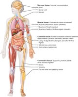
Four Basic Tissue Types: Epithelial, Connective, Muscle, and Nervous tissue.
Specialization: Individual body cells are specialized for specific functions.
Preparation of Tissue Samples for Microscopy
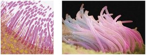
To study tissues, samples are fixed, sliced, and stained to enhance contrast for microscopic examination.
Fixed: Preserved with solvent.
Sectioned: Cut into thin slices for light or electron transmission.
Stained: Enhances contrast; light microscopy uses colored dyes, electron microscopy uses heavy metal salts.
Artifacts: Distortions that may detract from the true appearance of living tissues.
. '
Epithelial Tissue
Definition and Functions
Epithelial tissue consists of sheets of cells covering body surfaces or lining cavities. It is classified into two main forms: covering/lining epithelium and glandular epithelium.
Covering & Lining Epithelium: Skin, lining of urogenital, digestive, and respiratory systems.
Glandular Epithelium: Forms glands such as salivary glands.
Main Functions: Protection, absorption, filtration, excretion, secretion, and sensory reception.
Special Characteristics of Epithelial Tissue
Epithelial tissues possess unique features that distinguish them from other tissue types.
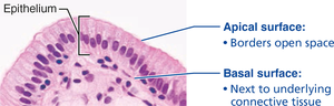
Polarity: Cells have distinct apical (top) and basal (bottom) surfaces.
Specialized Contacts: Tight junctions and desmosomes bind adjacent cells.
Supported by Connective Tissue: Basement membrane reinforces and defines boundaries.
Avascular but Innervated: No blood vessels, but supplied by nerve fibers.
Regeneration: High capacity for renewal, especially after damage.
Classification of Epithelial Tissue
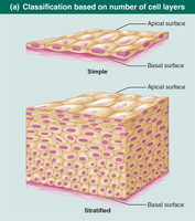
Epithelial tissues are classified based on the number of cell layers and cell shape.
Number of Layers: Simple (single layer) or Stratified (multiple layers).
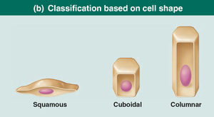
Cell Shape: Squamous (flat), Cuboidal (cube-like), Columnar (tall).
Stratified Epithelia: Named according to the shape of cells in the apical layer.
Types of Simple Epithelia
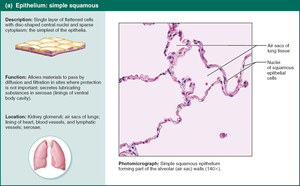
Simple Squamous Epithelium
Single layer of flattened cells, ideal for rapid diffusion and filtration.
Function: Allows materials to pass by diffusion and filtration.
Location: Kidney glomeruli, air sacs of lungs, lining of heart, blood vessels, lymphatic vessels.
Special Types: Endothelium (vessels, heart), Mesothelium (serous membranes).
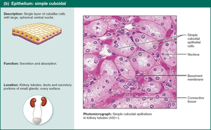
Simple Cuboidal Epithelium
Single layer of cube-shaped cells, involved in secretion and absorption.
Function: Secretion and absorption.
Location: Kidney tubules, ducts, and secretory portions of small glands.
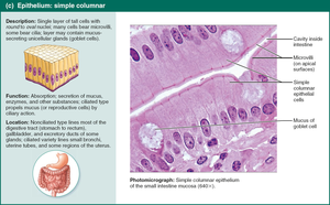
Simple Columnar Epithelium
Single layer of tall cells, often with microvilli or cilia, and goblet cells for mucus secretion.
Function: Absorption, secretion of mucus, enzymes, and other substances.
Location: Digestive tract, gallbladder, ducts of glands, bronchi, uterine tubes.
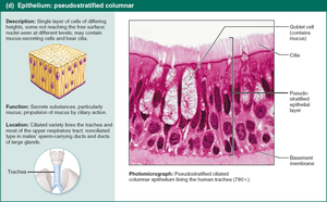
Pseudostratified Columnar Epithelium
Appears multi-layered but is actually a single layer; often ciliated and contains goblet cells.
Function: Secretion of mucus, movement of mucus by ciliary action.
Location: Upper respiratory tract, ducts of large glands, tubules in testes.
Connective Tissue
Overview and Functions
Connective tissue is the most abundant and widely distributed tissue type, serving functions such as binding, support, protection, insulation, storage, and transport.
Four Main Classes: Connective tissue proper, cartilage, bone, blood.
Extracellular Matrix: Non-living material that supports cells.
Common Origin: All connective tissues arise from mesenchyme.
Structural Elements of Connective Tissue
Ground Substance: Gel-like material with interstitial fluid, cell adhesion proteins, and proteoglycans.
Fibers: Collagen (strength), Elastic (stretch/recoil), Reticular (support networks).
Cells: Immature "-blast" cells (secrete matrix), mature "-cyte" cells (maintain matrix), adipocytes, leukocytes, mast cells, macrophages.
Types of Connective Tissue Proper
Loose Connective Tissue: Areolar, Adipose, Reticular.
Dense Connective Tissue: Dense regular, Dense irregular, Elastic.
Muscle Tissue
Types and Functions
Muscle tissue is responsible for movement and is highly vascularized. It contains myofilaments (actin and myosin) for contraction.
Skeletal Muscle: Voluntary, striated, multinucleated, attached to bones.
Cardiac Muscle: Involuntary, striated, one nucleus, intercalated discs, found in heart.
Smooth Muscle: Involuntary, non-striated, spindle-shaped, one nucleus, found in walls of hollow organs.
Nervous Tissue
Structure and Function
Nervous tissue regulates and controls body functions, consisting of neurons and glial cells.
Neurons: Respond to stimuli, transmit electrical signals via dendrites and axons.
Glial Cells: Support, insulate, and protect neurons.
Membranes: Covering and Lining
Types of Membranes
Membranes are organs composed of more than one tissue type, covering and lining body surfaces and cavities.
Cutaneous Membranes: Skin; keratinized stratified squamous epithelium attached to connective tissue.
Mucous Membranes: Line moist body cavities open to exterior; may secrete mucus.
Serous Membranes: Line closed ventral body cavities; constructed from mesothelium and areolar connective tissue.
Tissue Repair
Repair Mechanisms
Tissue repair occurs via regeneration (restoring original function) or fibrosis (replacement with connective tissue, loss of function).
Step 1: Inflammation sets the stage.
Step 2: Organization restores blood supply.
Step 3: Regeneration and fibrosis effect permanent repair.
Regenerative Capacity of Different Tissues
High Capacity: Epithelial, bone, areolar, dense irregular, blood-forming tissue.
Moderate Capacity: Smooth muscle, dense regular connective tissue.
Low/No Capacity: Cardiac muscle, nervous tissue of brain and spinal cord.
Developmental Aspects of Tissues
Tissues function well through youth and middle age, but efficiency declines with age. Epithelia thin, repair is less efficient, and risk of cancer increases due to DNA mutations.
