 뒤로
뒤로Chapter 4: Tissue – The Living Fabric (Anatomy & Physiology Study Notes)
스터디 가이드 - 스마트 노트
 자료에 맞춘 맞춤형 노트, 핵심 정의, 예시, 맥락을 확장해 제공합니다.
Tissue: The Living Fabric
Introduction to Tissues
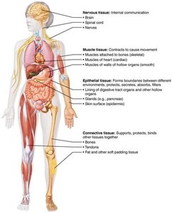
Tissues are groups of cells with similar structure and function, forming the fundamental building blocks of the human body. Understanding tissue types is essential for monitoring health and diagnosing conditions such as bedsores and cancer.
Histology: The study of tissues.
Four basic tissue types: Epithelial, connective, muscle, and nervous tissue.
Microscopy of Human Tissue
Preparation and Observation
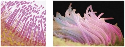
Microscopy is essential for studying tissues. Tissue samples must be fixed, sectioned, and stained to be viewed under a microscope. Light microscopy uses colored dyes, while electron microscopy uses heavy metal coatings.
Fixation: Preserves tissue structure.
Sectioning: Thin slices allow light/electron transmission.
Staining: Enhances contrast; may introduce artifacts.
Epithelial Tissue
Definition and Functions
Epithelial tissue forms sheets of cells covering surfaces and lining cavities. It is involved in protection, absorption, filtration, excretion, secretion, and sensory reception.
Covering and lining epithelia: External/internal surfaces (e.g., skin).
Glandular epithelia: Secretory tissue in glands (e.g., salivary glands).
Special Characteristics of Epithelial Tissue
Epithelial tissue is distinguished by five key characteristics:
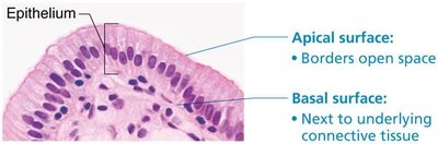
Polarity: Apical (top) and basal (bottom) surfaces differ in structure and function.
Specialized contacts: Tight junctions and desmosomes bind cells together.
Supported by connective tissue: Basement membrane reinforces and defines boundaries.
Avascular but innervated: No blood vessels; nourished by diffusion; supplied by nerves.
Regeneration: High capacity for renewal, especially after damage.
Classification of Epithelia
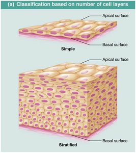
Epithelial tissues are classified by cell layers and cell shape:
Cell layers: Simple (one layer) or stratified (multiple layers).
Cell shape: Squamous (flat), cuboidal (cube-like), columnar (tall).
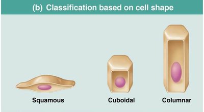
Types of Epithelial Tissue
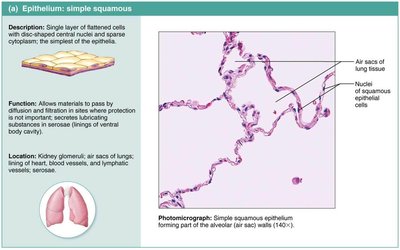
Simple squamous: Rapid diffusion; found in kidneys, lungs, blood vessels.
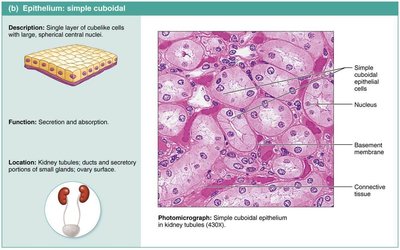
Simple cuboidal: Secretion and absorption; found in kidney tubules, glands.
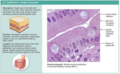
Simple columnar: Absorption, secretion; found in digestive tract, gallbladder, uterine tubes.
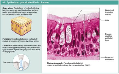
Pseudostratified columnar: Secretion and movement of mucus; found in respiratory tract.
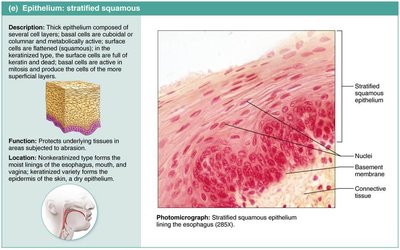
Stratified squamous: Protection; found in skin, mouth, esophagus.
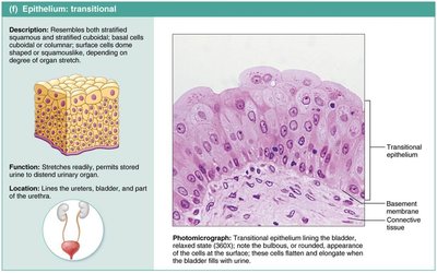
Transitional: Stretches; found in bladder, ureters, urethra.
Glandular Epithelia
Glands are cells or groups of cells that secrete aqueous fluids. They are classified by site of product release (endocrine or exocrine) and number of cells (unicellular or multicellular).
Endocrine glands: Ductless; secrete hormones into interstitial fluid and blood.
Exocrine glands: Secrete products into ducts; include sweat, oil, and salivary glands.
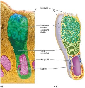
Unicellular exocrine glands: Goblet cells produce mucin.
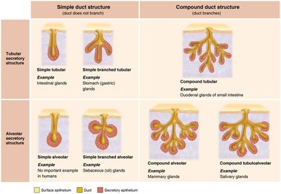
Multicellular exocrine glands: Composed of duct and secretory unit; classified by structure and mode of secretion.
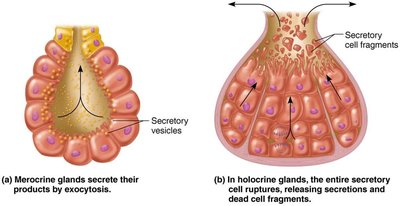
Modes of secretion: Merocrine (exocytosis), holocrine (rupture), apocrine (apex ruptures).
Covering and Lining Membranes
Membranes are composed of epithelial and connective tissues and include cutaneous, mucous, and serous membranes.
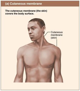
Cutaneous membrane: Skin; covers body surface.
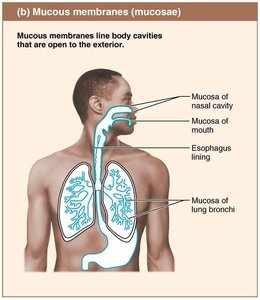
Mucous membrane: Lines body cavities open to exterior (e.g., respiratory, digestive).
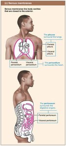
Serous membrane: Lines closed body cavities; produces serous fluid.
Connective Tissue
Definition and Functions
Connective tissue is the most abundant tissue, providing support, protection, insulation, storage, and transport. It is classified into four main classes: connective tissue proper, cartilage, bone, and blood.
Common characteristics: Mesenchymal origin, varying vascularity, extracellular matrix (ECM).
Comparison of Classes of Connective Tissues
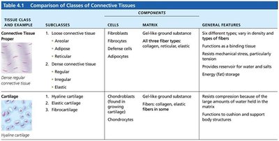
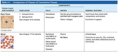
Connective tissues are compared based on their subclasses, cells, matrix, and general features.
Tissue Class | Subclasses | Cells | Matrix | General Features |
|---|---|---|---|---|
Connective Tissue Proper | Loose (areolar, adipose, reticular); Dense (regular, irregular, elastic) | Fibroblasts, adipocytes | Gel-like ground substance; collagen, elastic, reticular fibers | Binding, support, energy storage |
Cartilage | Hyaline, elastic, fibrocartilage | Chondroblasts, chondrocytes | Gel-like ground substance; collagen, elastic fibers | Resists compression, cushions |
Bone | Compact, spongy | Osteoblasts, osteocytes | Gel-like ground substance calcified with inorganic salts; collagen fibers | Support, protection |
Blood | --- | Red and white blood cells, platelets | Plasma (fluid); no fibers | Transport of substances |
Structural Elements of Connective Tissue
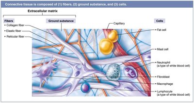
Connective tissue consists of ground substance, fibers, and cells. The composition and arrangement of these elements vary among tissue types.
Ground substance: Gel-like material; contains interstitial fluid, cell adhesion proteins, proteoglycans.
Fibers: Collagen (strength), elastic (stretch/recoil), reticular (support/give).
Cells: "Blast" (immature, matrix-secreting) and "cyte" (mature, maintenance) cells; fat cells, white blood cells, mast cells, macrophages.
Types of Connective Tissue Proper
Areolar: Supports and binds; universal packing material.
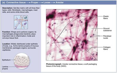
Adipose: Nutrient storage, insulation, shock absorption.
Reticular: Supports blood cells in lymph nodes, spleen, bone marrow.
Dense regular: High tensile strength; tendons, ligaments.
Dense irregular: Thicker, irregular bundles; dermis, joint capsules.
Elastic: Stretch/recoil; arteries, vertebral ligaments.
Cartilage
Hyaline: Most abundant; tips of long bones, nose, trachea.
Elastic: More elastic fibers; ears, epiglottis.
Fibrocartilage: Strong; intervertebral discs, knee.
Bone
Bone (osseous tissue) supports and protects, stores fat, and synthesizes blood cells. It is richly vascularized and contains osteons as structural units.
Blood
Blood is a fluid connective tissue, consisting of cells in plasma. It transports nutrients, wastes, gases, and other substances.
Muscle Tissue
Types and Functions
Muscle tissue is highly vascularized and responsible for movement. Muscle cells contain myofilaments (actin and myosin) for contraction. There are three types:
Skeletal muscle: Voluntary, striated, multiple nuclei; moves bones.
Cardiac muscle: Involuntary, striated, one nucleus, intercalated discs; found in heart.
Smooth muscle: Involuntary, no striations, spindle-shaped, one nucleus; walls of hollow organs.
Nervous Tissue
Structure and Function
Nervous tissue is the main component of the nervous system (brain, spinal cord, nerves). It regulates and controls body functions and consists of neurons (impulse-generating) and supporting cells (insulate, protect).
Tissue Repair
Steps in Tissue Repair
Tissue repair occurs via regeneration (restores original function) or fibrosis (connective tissue replaces, function lost). The process involves three steps:
Inflammation: Release of chemicals, dilation of blood vessels, clotting.
Organization: Blood clot replaced by granulation tissue, regeneration begins, fibroblasts bridge gap.
Regeneration and fibrosis: Scab detaches, tissue matures, scar tissue forms.
Regenerative Capacity of Different Tissues
High capacity: Epithelial, bone, areolar, dense irregular, blood-forming tissue.
Moderate capacity: Smooth muscle, dense regular connective tissue.
Low/no capacity: Cardiac muscle, nervous tissue (brain, spinal cord).
Clinical Relevance
Scar tissue can impair organ function, block substances, interfere with muscle contraction, or nerve transmission. Scar adhesions may cause organs to adhere abnormally, leading to progressive organ failure.
