 뒤로
뒤로Chapter 4: Tissues – The Living Fabric (Connective, Muscle, and Nervous Tissues)
스터디 가이드 - 스마트 노트
 자료에 맞춘 맞춤형 노트, 핵심 정의, 예시, 맥락을 확장해 제공합니다.
Chapter 4: Tissues – The Living Fabric
Overview of Connective Tissue
Connective tissue is the most abundant and widely distributed of the primary tissue types in the human body. It plays essential roles in binding and supporting other tissues, protecting organs, insulating the body, storing energy reserves, and transporting substances such as blood.
Major Functions: Binding/support, protection, insulation, energy storage, and transport.
Main Classes: Connective tissue proper, cartilage, bone, and blood.
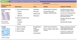
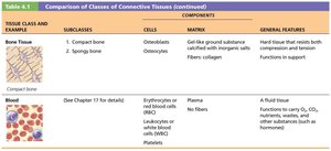
Main Classes of Connective Tissue
Tissue Class and Example | Subclasses | Cells | Matrix | General Features |
|---|---|---|---|---|
Connective Tissue Proper | Loose (areolar, adipose, reticular); Dense (regular, irregular, elastic) | Fibroblasts, fibrocytes, defense cells, adipocytes | Gel-like ground substance; all three fiber types (collagen, reticular, elastic) | Six different types; functions as a binding tissue; resists mechanical stress; provides water/nutrient reservoir; fat storage |
Cartilage | Hyaline, elastic, fibrocartilage | Chondroblasts, chondrocytes | Gel-like ground substance; fibers: collagen, elastic (in some) | Resists compression due to large amounts of water in matrix; cushions and supports body structures |
Bone Tissue | Compact, spongy | Osteoblasts, osteocytes | Gel-like ground substance calcified with inorganic salts; fibers: collagen | Hard tissue that resists both compression and tension; functions in support |
Blood | --- | Erythrocytes, leukocytes, platelets | Plasma (no fibers) | A fluid tissue; functions to carry CO2, nutrients, wastes, and other substances |
Common Characteristics of Connective Tissue
All connective tissues arise from mesenchyme (embryonic tissue).
They have varying degrees of vascularity (blood supply): cartilage is avascular, bone is highly vascularized.
Cells are suspended in an extracellular matrix (ECM) composed of protein-sugar mesh, allowing the tissue to bear weight, withstand tension, and endure physical abuse.
Structural Elements of Connective Tissue
All connective tissues share three main structural elements:
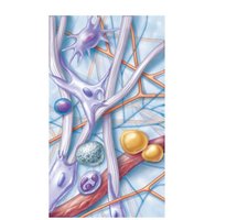
Ground Substance: Unstructured gel-like material that fills the space between cells and allows diffusion of nutrients and waste. Contains interstitial fluid, cell adhesion proteins, and proteoglycans (e.g., chondroitin sulfate, hyaluronic acid).
Fibers: Provide support and structure. Three types:
Collagen: Strongest and most abundant; provides tensile strength.
Elastic: Long, thin fibers that allow for stretch and recoil.
Reticular: Short, fine, highly branched fibers forming supportive networks.
Cells: Each class of connective tissue has a resident cell type:
"Blast" cells (immature, matrix-secreting): Fibroblasts (CT proper), Chondroblasts (cartilage), Osteoblasts (bone), Hematopoietic stem cells (blood).
"Cyte" cells (mature, maintain matrix): Fibrocytes, Chondrocytes, Osteocytes.
Other cells: Fat cells (store nutrients), White blood cells (defense), Mast cells (inflammation), Macrophages (phagocytosis).
Types of Connective Tissue
Connective Tissue Proper
This class includes all connective tissues except bone, cartilage, and blood. It is divided into two subclasses: loose and dense connective tissues.
Loose Connective Tissues
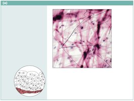
Areolar: Most widely distributed; supports and binds other tissues, holds body fluids, defends against infection, stores nutrients. Contains all three fiber types and various cells (fibroblasts, macrophages, mast cells, white blood cells).
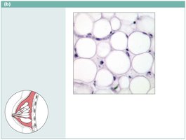
Adipose: Closely packed adipocytes (fat cells); stores energy, insulates, supports, and protects organs. Two types: white fat (energy storage, insulation, shock absorption) and brown fat (generates heat).
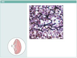
Reticular: Resembles areolar but with only reticular fibers; forms a soft internal skeleton (stroma) supporting blood cells in lymph nodes, spleen, and bone marrow.
Dense Connective Tissues
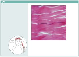
Dense Regular: Parallel collagen fibers; withstands tension in one direction; found in tendons and ligaments.
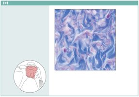
Dense Irregular: Thicker, irregularly arranged collagen bundles; resists tension from many directions; found in dermis, joint capsules, organ coverings.
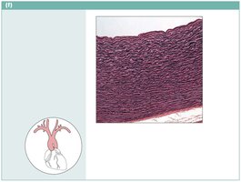
Elastic: High proportion of elastic fibers; allows tissue to recoil after stretching; found in walls of large arteries and certain ligaments.
Cartilage
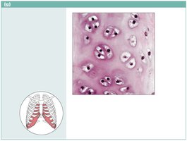
Cartilage is a tough yet flexible tissue that lacks nerve fibers and is avascular. It is composed of chondroblasts (during growth) and chondrocytes (in lacunae). The matrix is 80% water, with collagen fibers and proteoglycans.
Hyaline Cartilage: Most abundant; supports and reinforces; found at ends of long bones, nose, trachea, larynx, and ribs.
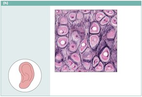
Elastic Cartilage: Similar to hyaline but with more elastic fibers; maintains shape and flexibility; found in ear and epiglottis.
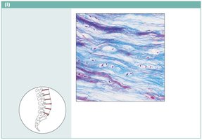
Fibrocartilage: Intermediate between hyaline cartilage and dense regular connective tissue; strong and resists compression; found in intervertebral discs, pubic symphysis, and knee discs.
Bone (Osseous Tissue)
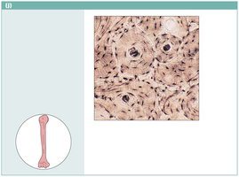
Bone tissue supports and protects body structures, stores fat, and synthesizes blood cells. It is highly vascularized and contains more collagen than cartilage. The matrix is calcified with inorganic salts. Osteoblasts produce the matrix, osteocytes maintain it, and both reside in lacunae. The basic structural unit is the osteon.
Blood
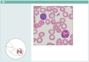
Blood is a fluid connective tissue consisting of cells (erythrocytes, leukocytes, platelets) suspended in plasma. It functions in the transport of gases, nutrients, wastes, and other substances throughout the body.
Muscle Tissue
Muscle tissue is highly vascularized and responsible for most types of body movement. Muscle cells contain myofilaments (actin and myosin) that enable contraction. There are three types of muscle tissue:
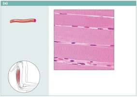
Skeletal Muscle: Voluntary, striated, multinucleate cells; attached to bones and responsible for movement.
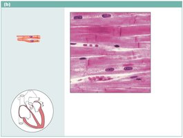
Cardiac Muscle: Involuntary, striated, usually uninucleate, branching cells connected by intercalated discs; found only in the heart.
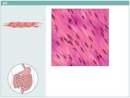
Smooth Muscle: Involuntary, non-striated, spindle-shaped cells with one nucleus; found in walls of hollow organs (except heart).
Nervous Tissue
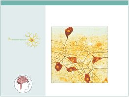
Nervous tissue is the main component of the nervous system (brain, spinal cord, nerves). It regulates and controls body functions. It consists of two main cell types:
Neurons: Specialized cells that generate and conduct nerve impulses.
Supporting Cells: Support, insulate, and protect neurons.
Covering and Lining Membranes
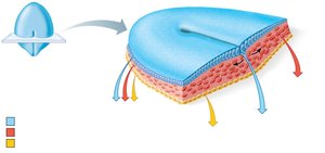
Membranes are composed of at least two primary tissue types: an epithelium bound to underlying connective tissue. There are three main types:
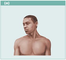
Cutaneous Membrane: The skin; a dry membrane composed of keratinized stratified squamous epithelium attached to a thick layer of connective tissue.
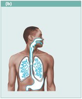
Mucous Membranes (Mucosae): Line body cavities open to the exterior (digestive, respiratory, urogenital tracts); moist membranes bathed by secretions; consist of an epithelial sheet over a layer of loose connective tissue (lamina propria).
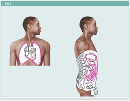
Serous Membranes (Serosae): Line closed ventral body cavities; composed of simple squamous epithelium (mesothelium) on areolar connective tissue; secrete serous fluid; include pleurae (lungs), pericardium (heart), peritoneum (abdomen).
Tissue Repair
Tissue repair occurs when the body's barriers are compromised. The process involves inflammation, organization, and regeneration or fibrosis.
Steps in Tissue Repair
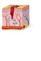
Inflammation: Inflammatory chemicals are released, blood vessels dilate and become more permeable, and clotting occurs, forming a scab.
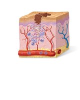
Organization: The blood clot is replaced by granulation tissue, restoring vascular supply. Fibroblasts produce collagen fibers, and macrophages remove debris. Surface epithelial cells multiply and migrate.
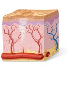
Regeneration and Fibrosis: The scab detaches, fibrous tissue matures, and the epithelium thickens, resulting in a fully regenerated epithelium with underlying scar tissue.
Regenerative Capacity of Different Tissues
High regenerative capacity: Epithelial tissues, bone, areolar connective tissue, dense irregular connective tissue, blood-forming tissue.
Moderate capacity: Smooth muscle, dense regular connective tissue.
Virtually no functional capacity: Cardiac muscle, nervous tissue in brain and spinal cord.
Developmental Aspects of Tissues
Primary germ layers: Ectoderm, mesoderm, endoderm (from superficial to deep).
Nerve tissue arises from ectoderm; muscle and connective tissues from mesoderm; epithelial tissues from all three layers.
Tissues function well through youth and middle age; aging leads to thinning epithelia, less efficient repair, atrophy of bone/muscle/nervous tissue, and increased cancer risk due to DNA mutations.
Additional info: This summary integrates textbook content, tables, and histological images to provide a comprehensive overview of connective, muscle, and nervous tissues, as well as tissue repair and development, suitable for college-level Anatomy & Physiology students.
