 뒤로
뒤로Chapter 4: Tissue—The Living Fabric (Anatomy & Physiology Study Notes)
스터디 가이드 - 스마트 노트
 자료에 맞춘 맞춤형 노트, 핵심 정의, 예시, 맥락을 확장해 제공합니다.
Tissue: The Living Fabric
Introduction to Tissues
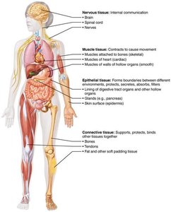
Tissues are groups of cells similar in structure that perform common or related functions. The study of tissues is called histology. There are four basic tissue types in the human body: epithelial, connective, muscle, and nervous tissue. Each type has specialized roles essential for maintaining homeostasis.
Epithelial tissue: Covers body surfaces, lines cavities, and forms glands.
Connective tissue: Supports, protects, and binds other tissues.
Muscle tissue: Responsible for movement.
Nervous tissue: Enables internal communication via electrical impulses.
Microscopy of Human Tissue
Preparation and Staining
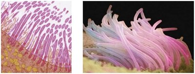
To study tissues under a microscope, they must be:
Fixed: Preserved with a solvent.
Sectioned: Cut into thin slices for light or electron transmission.
Stained: Enhanced contrast using dyes (light microscopy) or heavy metals (electron microscopy).
Epithelial Tissue
General Characteristics
Epithelial tissue (epithelium) is a sheet of cells that covers body surfaces or lines cavities. It exists in two main forms:
Covering and lining epithelia: On external and internal surfaces (e.g., skin).
Glandular epithelia: Secretory tissue in glands (e.g., salivary glands).
Main functions: protection, absorption, filtration, excretion, secretion, and sensory reception.
Special Characteristics of Epithelial Tissue
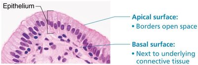
Polarity: Cells have an apical (top) and basal (bottom) surface, each with distinct structures and functions.
Specialized contacts: Cells are closely joined by tight junctions and desmosomes.
Supported by connective tissue: All epithelial sheets rest on a basement membrane (basal and reticular lamina).
Avascular but innervated: No blood vessels, but supplied by nerves; nutrients diffuse from underlying tissues.
Regeneration: High capacity for renewal, especially in areas exposed to friction or hostile environments.
Classification of Epithelia
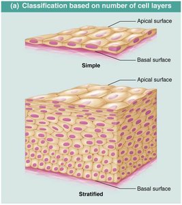
Epithelia are classified by the number of cell layers and cell shape:
Layers: Simple (one layer) or stratified (multiple layers).
Shapes: Squamous (flat), cuboidal (cube-like), columnar (tall).
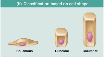
Types of Epithelial Tissue
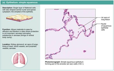
Simple Squamous Epithelium: Single layer of flat cells; allows diffusion/filtration (e.g., air sacs of lungs, lining of blood vessels).
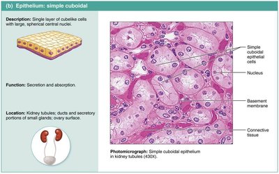
Simple Cuboidal Epithelium: Single layer of cube-shaped cells; secretion and absorption (e.g., kidney tubules, small glands).
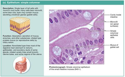
Simple Columnar Epithelium: Single layer of tall cells; absorption, secretion of mucus/enzymes (e.g., digestive tract, uterine tubes).
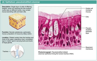
Pseudostratified Columnar Epithelium: Appears multi-layered but is single-layered; secretion and movement of mucus (e.g., trachea).
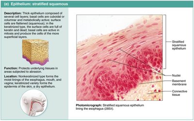
Stratified Squamous Epithelium: Multiple layers; protects underlying tissues (e.g., skin, mouth, esophagus).
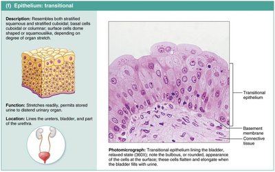
Transitional Epithelium: Resembles both stratified squamous and cuboidal; stretches (e.g., bladder, ureters).
Glandular Epithelia
A gland consists of one or more cells that secrete an aqueous fluid (secretion). Glands are classified by:
Site of product release: Endocrine (into blood/lymph) or exocrine (onto surfaces or into ducts).
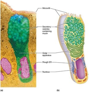
Number of cells: Unicellular (e.g., goblet cells) or multicellular (e.g., salivary glands).
Types of Multicellular Exocrine Glands
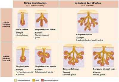
Classified by structure (simple/compound, tubular/alveolar) and mode of secretion:
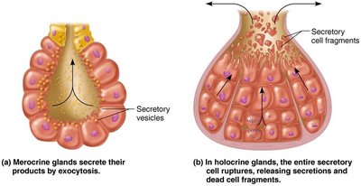
Merocrine: Secrete by exocytosis (e.g., sweat glands).
Holocrine: Accumulate products, then rupture (e.g., sebaceous glands).
Apocrine: Apex ruptures (controversial in humans).
Covering and Lining Membranes
Three types of membranes combine epithelial and connective tissues:
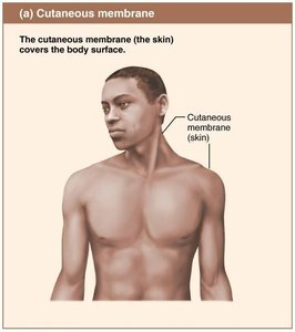
Cutaneous membrane: The skin; covers the body surface.
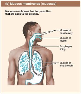
Mucous membranes: Line body cavities open to the exterior (e.g., digestive, respiratory tracts).
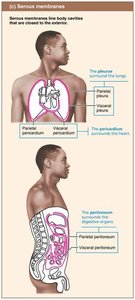
Serous membranes: Line closed ventral body cavities (e.g., pleura, pericardium, peritoneum).
Connective Tissue
General Characteristics
Connective tissue is the most abundant and widely distributed tissue type. Its major functions include binding/support, protection, insulation, energy storage, and transport (blood). Four main classes:
Connective tissue proper
Cartilage
Bone
Blood
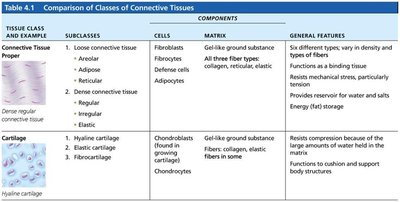
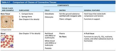
Comparison of Classes of Connective Tissues
Tissue Class | Subclasses | Cells | Matrix | General Features |
|---|---|---|---|---|
Connective Tissue Proper | Loose (areolar, adipose, reticular); Dense (regular, irregular, elastic) | Fibroblasts, fibrocytes, defense cells, adipocytes | Gel-like ground substance; all three fiber types | Binding tissue, resists tension, energy storage |
Cartilage | Hyaline, elastic, fibrocartilage | Chondroblasts, chondrocytes | Gel-like ground substance; collagen, elastic fibers | Resists compression, cushions body structures |
Bone | Compact, spongy | Osteoblasts, osteocytes | Gel-like ground substance calcified with inorganic salts; collagen fibers | Hard tissue, support, resists tension/compression |
Blood | --- | RBCs, WBCs, platelets | Plasma (no fibers) | Fluid tissue, transport of substances |
Common Characteristics of Connective Tissue
All arise from mesenchyme (embryonic tissue).
Varying degrees of vascularity (cartilage is avascular, bone is highly vascularized).
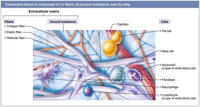
Cells are suspended in an extracellular matrix (ECM) of fibers and ground substance.
Structural Elements of Connective Tissue
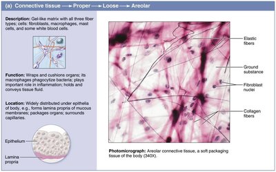
Ground substance: Unstructured material filling space between cells; includes interstitial fluid, cell adhesion proteins, proteoglycans.
Fibers: Collagen (strength), elastic (stretch/recoil), reticular (supportive mesh).
Cells: "Blast" (immature, matrix-secreting) and "cyte" (mature, maintenance) forms; also fat cells, WBCs, mast cells, macrophages.
Types of Connective Tissue Proper
Loose connective tissue: Areolar, adipose, reticular.
Dense connective tissue: Dense regular, dense irregular, elastic.
Muscle Tissue
General Characteristics
Muscle tissue is highly vascularized and responsible for movement. Muscle cells contain myofilaments (actin and myosin) for contraction. Three types:
Skeletal muscle: Voluntary, striated, multinucleate, attached to bones.
Cardiac muscle: Involuntary, striated, one nucleus, intercalated discs, found in heart.
Smooth muscle: Involuntary, non-striated, spindle-shaped, one nucleus, found in walls of hollow organs.
Nervous Tissue
General Characteristics
Nervous tissue is the main component of the nervous system (brain, spinal cord, nerves). It regulates and controls body functions. Two main cell types:
Neurons: Generate and conduct nerve impulses.
Supporting cells: Support, insulate, and protect neurons.
Tissue Repair
Steps in Tissue Repair
When tissue is damaged, repair occurs via two main processes:
Regeneration: Replacement with the same tissue type, restoring function.
Fibrosis: Replacement with connective tissue (scar), function may be lost.
Inflammation: Release of chemicals, dilation of blood vessels, increased permeability, clotting.
Organization: Blood clot replaced by granulation tissue, epithelium regenerates, fibroblasts bridge gap.
Regeneration/Fibrosis: Scab detaches, tissue matures, scar may remain.
Regenerative Capacity of Tissues
High: Epithelial, bone, areolar, dense irregular, blood-forming tissue.
Moderate: Smooth muscle, dense regular connective tissue.
Low/None: Cardiac muscle, nervous tissue in brain/spinal cord.
Clinical Relevance
Scar tissue in organs (especially heart) can impair function, block movement, or cause organ failure.
