 뒤로
뒤로Chapter 5: The Skeletal System – Structure, Function, and Articulations
스터디 가이드 - 스마트 노트
 자료에 맞춘 맞춤형 노트, 핵심 정의, 예시, 맥락을 확장해 제공합니다.
The Skeletal System
Functions of the Skeletal System
The skeletal system is a dynamic organ system that provides the framework for the human body and supports a variety of essential physiological functions.
Support: The skeleton forms the structural framework for the body, supporting soft tissues and providing attachment points for muscles.
Protection: Bones protect vital organs, such as the brain (skull), heart and lungs (rib cage), and spinal cord (vertebrae).
Movement: Bones act as levers and points of attachment for muscles, enabling movement when muscles contract.
Mineral Storage: Bones store minerals, primarily calcium and phosphorus, which can be released into the bloodstream as needed.
Fat Storage: Yellow bone marrow stores triglycerides, serving as an energy reserve.
Blood Cell Formation: Red bone marrow is the site of hematopoiesis, the production of blood cells.
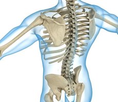
Classification and Structure of Bones
Types of Bones
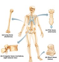
Bones are classified according to their shapes and locations in the body, which relate to their functions.
Long Bones: Longer than they are wide (e.g., femur, humerus); primarily function as levers.
Short Bones: Nearly equal in length and width (e.g., carpals, tarsals); provide stability and support.
Flat Bones: Thin, flattened, and often curved (e.g., sternum, ribs, skull bones); protect internal organs and provide surfaces for muscle attachment.
Irregular Bones: Complex shapes that do not fit other categories (e.g., vertebrae, some facial bones).
Sesamoid Bones: Small, round bones embedded within tendons (e.g., patella); protect tendons from stress and wear.
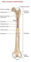
Gross Structure of a Long Bone
Long bones have a characteristic structure that supports their function in movement and weight-bearing.
Epiphysis: The expanded ends of the bone, covered with articular cartilage for joint movement.
Diaphysis: The shaft or central part of the bone, composed mainly of compact bone.
Periosteum: A dense irregular collagenous connective tissue membrane covering the outer surface of the bone, containing nerves and blood vessels.
Compact Bone: Dense, hard outer layer that provides strength and protection.
Spongy Bone (Cancellous Bone): Porous, lighter bone found mainly in the epiphyses; contains red marrow.
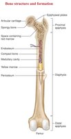
Internal Bone Features
Articular Cartilage: Hyaline cartilage covering joint surfaces, reducing friction and absorbing shock.
Medullary Cavity: Central cavity within the diaphysis, housing yellow marrow (fat storage) in adults and red marrow (blood cell production) in children.
Red Marrow: Site of hematopoiesis (blood cell formation).
Yellow Marrow: Stores triglycerides (fat).
Microscopic Structure of Bone Tissue
Osteon and Bone Cells
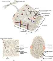
The functional unit of compact bone is the osteon, which is organized to provide strength and facilitate nutrient delivery.
Osteon (Haversian System): Cylindrical structures containing a central canal surrounded by concentric lamellae.
Central Canal: Contains blood vessels and nerves.
Lamellae: Rings of calcified matrix.
Canaliculi: Small channels connecting osteocytes for nutrient and waste exchange.
Trabeculae: Lattice-like structures in spongy bone, providing support and housing marrow.
Osteoblasts: Bone-forming cells that become osteocytes when trapped in matrix.
Osteoclasts: Bone-resorbing cells involved in bone remodeling.
Bone Fractures
Classification of Bone Fractures
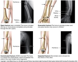
Bone fractures are classified based on the nature and extent of the break. Understanding these types is essential for diagnosis and treatment.
Type of Fracture | Description |
|---|---|
Open (Compound) | Bone breaks through the skin |
Greenstick | Bone is partially broken and partially bent (common in children) |
Comminuted | Bone is shattered into three or more pieces |
Impacted | One bone fragment is driven into another |
Bones of the Skeleton
Skull
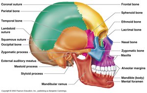
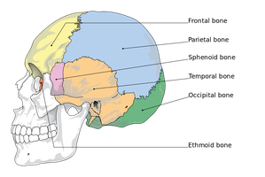
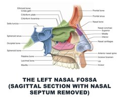
The skull protects the brain and forms the structure of the face. It is composed of cranial and facial bones.
Cranial Bones: Enclose and protect the brain (e.g., frontal, parietal, temporal, occipital, sphenoid, ethmoid).
Facial Bones: Form the structure of the face (e.g., maxilla, mandible, zygomatic, nasal, lacrimal, palatine, inferior nasal concha, vomer).
Paranasal Sinuses
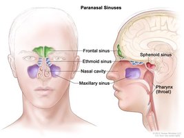
Paranasal sinuses are air-filled spaces within certain skull bones that lighten the skull and enhance voice resonance.
Frontal, Ethmoid, Sphenoid, and Maxillary Sinuses: Located within the respective bones; lined with mucous membranes.
Vertebral Column
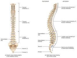
The vertebral column (spine) supports the body, protects the spinal cord, and provides attachment points for ribs and muscles.
Cervical (7), Thoracic (12), Lumbar (5), Sacrum, Coccyx: Regions of the vertebral column, each with unique features.
Spinal Disorders & Injuries
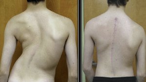
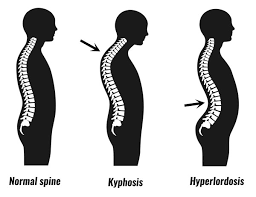
Scoliosis: Lateral curvature of the spine.
Kyphosis: Excessive posterior curvature (hunchback).
Lordosis: Excessive anterior curvature (swayback).
Herniated Discs: Protrusion of intervertebral disc, causing nerve compression.
Rib Cage
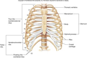
The rib cage protects the heart and lungs and supports the upper body.
True Ribs (1-7): Attach directly to the sternum.
False Ribs (8-12): Attach indirectly or not at all to the sternum.
Floating Ribs (11-12): Do not attach to the sternum.
Pectoral Girdle and Upper Limb
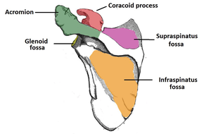
Scapula: Shoulder blade; provides attachment for arm muscles.
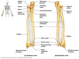
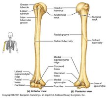
Humerus, Radius, Ulna: Bones of the arm and forearm.
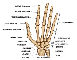
Carpals, Metacarpals, Phalanges: Bones of the wrist and hand.
Pelvic Girdle and Lower Limb
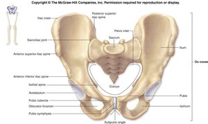
Pelvis: Supports the weight of the upper body and protects pelvic organs.
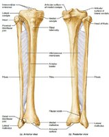
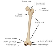
Femur, Tibia, Fibula: Bones of the thigh and leg.
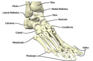
Tarsals, Metatarsals, Phalanges: Bones of the ankle and foot.
Articulations (Joints)
Classification of Joints
Joints are classified by their structure and function, which determine their range of movement and stability.
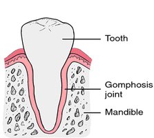
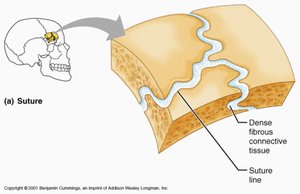
Fibrous Joints: Bones joined by dense connective tissue; mostly immovable (e.g., sutures of the skull, gomphosis of teeth, syndesmosis between parallel bones).
Cartilaginous Joints: Bones joined by cartilage; allow limited movement (e.g., synchondroses, symphyses).
Synovial Joints: Freely moveable joints with a synovial cavity (e.g., knee, shoulder, hip).
Fibrous Joints
Sutures: Immovable joints between skull bones; replaced by bone with age.
Gomphosis: Peg-in-socket joint (e.g., tooth in alveolar socket).
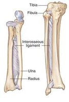
Syndesmosis: Bones connected by an interosseous membrane (e.g., between radius and ulna).
Cartilaginous Joints
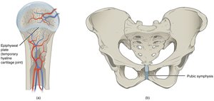
Synchondroses: Bones united by hyaline cartilage (e.g., epiphyseal plate, costal cartilage of ribs).
Symphyses: Bones joined by fibrocartilage (e.g., intervertebral discs, pubic symphysis); allow limited movement.
Synovial Joints
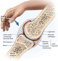
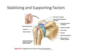
Articular Cartilage: Covers bone surfaces at joints, reducing friction.
Articular Capsule: Encloses the joint cavity, lined with synovial membrane.
Joint Cavity: Contains synovial fluid for lubrication.
Reinforcing Ligaments: Strengthen and stabilize the joint.
Types of Synovial Joints by Structure
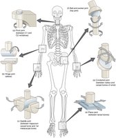
Plane: Gliding movements (e.g., intercarpal joints).
Hinge: Flexion and extension (e.g., elbow, knee).
Pivot: Rotation around a single axis (e.g., atlas and axis of the neck).
Condylar: Movement in two planes (e.g., wrist).
Saddle: Allows movement in two planes (e.g., thumb joint).
Ball-and-Socket: Multiaxial movement (e.g., shoulder, hip).
Types of Movement at Synovial Joints
Gliding: Sliding movements between flat surfaces.
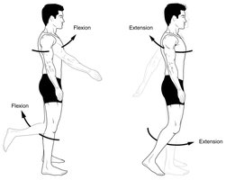
Flexion vs Extension: Decreasing vs increasing the angle between bones.
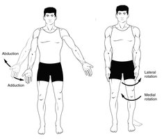
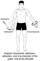
Abduction vs Adduction: Movement away from vs toward the midline.
Circumduction: Circular movement of a limb.
Rotation: Movement around a longitudinal axis.
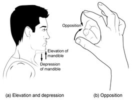
Opposition vs Reposition: Movement of the thumb to touch other fingers vs returning to anatomical position.
Depression vs Elevation: Lowering vs raising a body part.
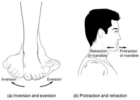
Protraction vs Retraction: Moving a body part forward vs backward.
Inversion vs Eversion: Turning the sole of the foot inward vs outward.
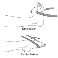
Dorsiflexion vs Plantarflexion: Raising the foot upward vs pointing it downward.
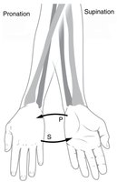
Supination vs Pronation: Rotating the forearm to turn the palm up vs down.
Stabilizing Synovial Joints
Ligaments: Connect bone to bone, providing stability.
Tendons: Connect muscle to bone, contributing to joint stability.
Bursae: Fluid-filled sacs that reduce friction between moving structures.
Tendon Sheaths: Tubular bursae that surround tendons.
Major Synovial Joints
Shoulder (Glenohumeral Joint): Ball-and-socket joint with a labrum (cartilage) lining the socket for stability.
Hip (Coxal Joint): Ball-and-socket joint with strong ligaments and a deep socket for weight-bearing stability.
Knee: Largest and most complex joint; hinge joint with menisci and multiple ligaments for stability.
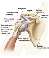
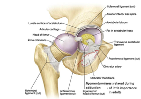
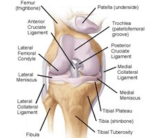
Additional info: This guide covers the essential structure and function of the skeletal system, including bone types, gross and microscopic anatomy, major bones of the body, joint classification, and common movements. Disorders such as scoliosis, kyphosis, and lordosis are also introduced for clinical context.
