 뒤로
뒤로Classification of Tissues: Epithelial Tissue Structure and Function
스터디 가이드 - 스마트 노트
 자료에 맞춘 맞춤형 노트, 핵심 정의, 예시, 맥락을 확장해 제공합니다.
Classification of Tissues
Overview of Tissue Types
Tissues are groups of cells that are similar in structure and function. In human anatomy and physiology, tissues are classified into four primary types: epithelial, connective, muscle, and nervous tissue. Each type has distinct roles in the body.
Epithelial Tissue: Covers and lines surfaces; forms glands.
Connective Tissue: Provides support; includes bone, ligaments, and fat.
Muscle Tissue: Responsible for movement.
Nervous Tissue: Controls and coordinates body activities; found in the brain, nerves, and spinal cord.

Epithelial Tissue
General Characteristics
Epithelial tissue consists of layers of cells that cover internal or external surfaces and form glands. It is specialized for protection, absorption, filtration, and secretion.
Continuous Sheets: Epithelial cells fit closely together, forming continuous layers.
Apical Surface: The top surface of epithelial cells faces an open space (lumen).
Basement Membrane: The underside of epithelial cells is anchored to connective tissue.
Avascularity: Epithelial tissue lacks blood vessels and is nourished by underlying connective tissue.
Regeneration: Epithelial cells regenerate and repair quickly.
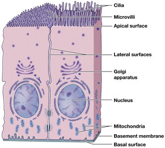
Functions of Epithelial Tissue
Protection: Forms barriers against physical, chemical, and biological harm (e.g., skin protects from sunlight and bacteria).
Absorption: Specialized for nutrient uptake (e.g., lining of the small intestine).
Filtration: Filters substances (e.g., kidney tubules filter blood plasma).
Secretion: Produces and releases substances (e.g., sweat, oil, digestive enzymes, mucus).
Classification of Epithelial Tissue
By Cell Shape and Layers
Epithelial tissues are classified based on the shape of their cells and the number of cell layers.
Cell Shape:
Squamous: Flattened, scale-like cells.
Cuboidal: Cube-shaped cells.
Columnar: Tall, column-like cells.
Transitional: Cells that change shape (found in organs that stretch).
Cell Layers:
Simple: One layer of cells.
Stratified: Multiple layers of cells.
Pseudostratified: Appears multilayered but is a single layer with nuclei at different levels.
Types of Epithelial Tissue
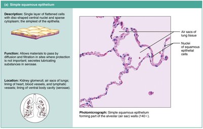
Simple Squamous Epithelium
Simple squamous epithelium consists of a single layer of flattened cells. It is specialized for absorption and filtration, but not for protection due to its thinness.
Location: Air sacs of lungs, lining of heart and blood vessels (endothelium), lining of body cavities (mesothelium).
Function: Allows diffusion and filtration of materials.
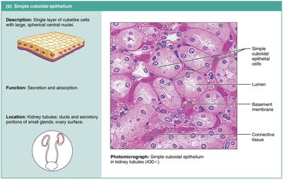
Simple Cuboidal Epithelium
Simple cuboidal epithelium is composed of a single layer of cube-shaped cells. It is involved in secretion and absorption.
Location: Kidney tubules, ducts and secretory portions of small glands, ovary surface.
Function: Secretion and absorption.
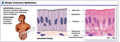
Simple Columnar Epithelium
Simple columnar epithelium consists of a single layer of tall, column-like cells with nuclei aligned at the same level. It is specialized for absorption, protection, and secretion.
Special Features: May have microvilli (increase surface area) and goblet cells (secrete mucus).
Location: Lining of the digestive tract (stomach to rectum), gallbladder, uterine tubes.
Function: Absorption, secretion of mucus and enzymes, protection.
Pseudostratified Columnar Epithelium
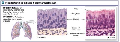
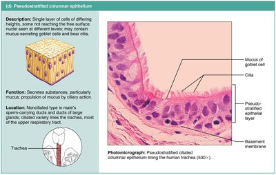
Pseudostratified columnar epithelium appears to have multiple layers due to nuclei at different levels, but all cells rest on the basement membrane. It often contains cilia and goblet cells.
Location: Lining of respiratory tract (trachea, bronchi), parts of male reproductive tract.
Function: Secretion (especially mucus), propulsion of mucus by ciliary action.
Stratified Squamous Epithelium
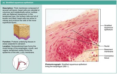
Stratified squamous epithelium is composed of several layers, with cuboidal or columnar cells at the base and flattened squamous cells at the surface. It is designed for protection against abrasion.
Location: Epidermis of skin (keratinized), lining of mouth, esophagus, and vagina (non-keratinized).
Function: Protects underlying tissues in areas subject to abrasion.
Special Feature: Keratinization in skin provides waterproofing and toughness.
Transitional Epithelium
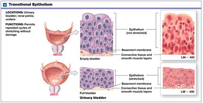
Transitional epithelium consists of multiple layers of cells that can change shape from cuboidal to squamous as the tissue stretches. This allows organs to expand and contract.
Location: Urinary bladder, ureters, and part of the urethra.
Function: Permits repeated cycles of stretching without damage.
Glandular Epithelium
Glands and Their Classification
Glands are composed of one or more cells that produce and secrete a specific product, such as hormones, enzymes, or oils. Glands are classified as endocrine or exocrine based on how they release their products.
Endocrine Glands: Ductless; secrete hormones directly into the bloodstream (e.g., thyroid, adrenal, pituitary glands).
Exocrine Glands: Have ducts; secrete products onto epithelial surfaces (e.g., sweat, oil, salivary, and mammary glands).
Structural Classification of Exocrine Glands
Exocrine glands are further classified by the structure of their ducts and the shape of their secretory units.
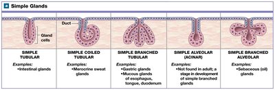
Simple Glands: Unbranched ducts.
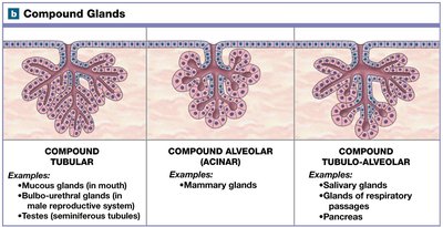
Compound Glands: Branched ducts.
Secretory Unit Shape:
Tubular: Tube-shaped.
Alveolar (Acinar): Flask- or sac-shaped.
Tubuloalveolar: Contains both tubular and alveolar regions.
Type | Examples |
|---|---|
Simple Tubular | Intestinal glands |
Simple Coiled Tubular | Merocrine sweat glands |
Simple Branched Tubular | Gastric glands, mucous glands |
Simple Alveolar | Not found in adults |
Simple Branched Alveolar | Sebaceous (oil) glands |
Compound Tubular | Mucous glands (mouth), seminiferous tubules (testes) |
Compound Alveolar | Mammary glands |
Compound Tubuloalveolar | Salivary glands, pancreas |
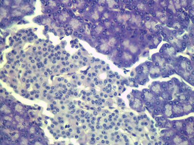
Exocrine and Endocrine Cells in the Pancreas
The pancreas contains both exocrine and endocrine cells. Exocrine cells secrete digestive enzymes into ducts, while endocrine cells release hormones (such as insulin and glucagon) directly into the bloodstream.
Additional info: The classification and structure of epithelial tissues are foundational for understanding organ function and pathology in human anatomy and physiology. Mastery of these concepts is essential for further study of tissue histology and organ systems.
