 뒤로
뒤로Cytology: Structure and Function of the Cell
스터디 가이드 - 스마트 노트
 자료에 맞춘 맞춤형 노트, 핵심 정의, 예시, 맥락을 확장해 제공합니다.
The Cell: Basic Unit of Life
Overview of Cell Types
The cell is the smallest living unit in the body and forms the foundation of all biological structure and function. Cells are classified based on the presence or absence of a membrane-bound nucleus and organelles.
Eukaryotic cells: Possess a membrane-bound nucleus and organelles. Found in protists, plants, fungi, and animals.
Prokaryotic cells: Lack a membrane-bound nucleus and organelles. Found in bacteria and archaea.
Cellular Structures and Organelles
Plasma Membrane
The plasma membrane is a dynamic, selectively permeable barrier composed of a fluid mosaic of lipids and proteins. It separates the intracellular environment from the extracellular space and regulates the movement of substances into and out of the cell.
Gatekeeper function: Allows small, hydrophobic molecules (e.g., CO2, O2) to pass directly through, while large or hydrophilic molecules (e.g., H2O, ions) require specialized transport mechanisms.
Nucleus
The nucleus serves as the control center of the cell, housing genetic material and coordinating cellular activities.
Nuclear envelope: Double lipid bilayer that encloses the nucleus.
Nuclear pores: Regulate the movement of proteins, RNA, and other molecules.
Nucleolus: Site of ribosomal RNA synthesis.
Chromatin: DNA wrapped around histone proteins, visible as grainy material during interphase.
Endoplasmic Reticulum (ER)
Rough ER (RER): Studded with ribosomes; synthesizes glycoproteins and secretory proteins. Ribosomes not bound to RER (polysomes) produce proteins for intracellular use.
Smooth ER (sER): Lacks ribosomes; synthesizes lipids, metabolizes carbohydrates, detoxifies drugs/toxins (abundant in liver), and stores calcium ions (important for muscle contraction).
Golgi Apparatus
The Golgi apparatus is a series of flattened membranous sacs that modify, sort, and package proteins and lipids received from the ER.
Packages products into secretory vesicles for lysosome formation, membrane renewal, or exocytosis.
Lysosomes
Lysosomes contain hydrolytic enzymes that digest macromolecules, bacteria, and damaged organelles. They are essential for autolysis and phagocytosis.
Mitochondria
Mitochondria are double-membrane-bound organelles responsible for ATP production via aerobic respiration. They are abundant in energy-demanding tissues such as skeletal muscle and are believed to have evolved from engulfed prokaryotes.
Produce free radicals as byproducts.
Can contribute to autolysis.
Cytoskeleton
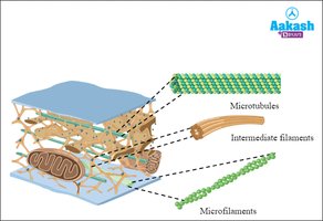
The cytoskeleton provides structural support, facilitates intracellular transport, and is involved in cell division and movement. It is composed of three main types of protein filaments:
Microtubules: Hollow rods made of tubulin; form the basis for organelle transport and the mitotic spindle.
Intermediate filaments: Provide mechanical support and maintain cell shape.
Microfilaments: Thin filaments composed of actin; involved in cell movement and shape changes.
Centrioles
Centrioles are cylindrical structures composed of microtubules. They organize the mitotic spindle during cell division and are found in pairs within the centrosome near the nucleus during interphase.
Cell Cycle and Mitosis
Interphase
Interphase is the period of cell growth and preparation for division, consisting of three stages:
G1 phase: Cell growth; synthesis of mRNA and proteins required for DNA and organelle replication.
S phase: DNA replication occurs.
G2 phase: Protein synthesis continues in preparation for mitosis.
Mitosis
Mitosis is the process by which a cell divides its nucleus and genetic material to produce two identical daughter cells. It consists of several stages:
Prophase
Nuclear envelope dissolves.
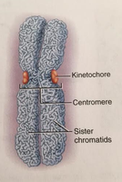
Chromatin condenses into visible chromosomes (paired sister chromatids joined at the centromere).
Centrioles replicate and migrate to opposite poles, forming the mitotic spindle apparatus.
Mitotic Spindle Apparatus
The mitotic spindle is composed of microtubules that attach to chromosomes via the kinetochore, a protein complex at the centromere. The spindle apparatus is essential for chromosome movement during mitosis.
Metaphase
Chromosomes align at the metaphase plate, an imaginary plane equidistant from the spindle poles.
Kinetochore microtubules attach to each chromatid.
Anaphase
Shortest stage of mitosis.
Sister chromatids separate and move toward opposite poles, pulled by the spindle apparatus.
The cell elongates to facilitate separation.
Telophase and Cytokinesis
Nuclear envelopes reform around the separated chromosomes.
Chromosomes begin to decondense.
Cytokinesis (division of the cytoplasm) occurs, often beginning in late anaphase, involving the formation of a cleavage furrow that pinches the cell into two.
Summary Table: Key Cell Structures and Functions
Structure | Main Function | Key Features |
|---|---|---|
Plasma Membrane | Selective barrier, communication | Lipid bilayer, proteins, fluid mosaic |
Nucleus | Genetic control center | Nuclear envelope, nucleolus, chromatin |
Rough ER | Protein synthesis | Ribosomes attached |
Smooth ER | Lipid synthesis, detoxification | No ribosomes, tubular |
Golgi Apparatus | Protein modification & packaging | Stacked sacs |
Lysosome | Digestion, autolysis | Hydrolytic enzymes |
Mitochondrion | ATP production | Double membrane, cristae |
Centrioles | Spindle formation | Microtubule-based |
Cytoskeleton | Support, movement | Microtubules, intermediate filaments, microfilaments |
