 뒤로
뒤로Cytoplasm, Organelles, and Cellular Processes: Structure and Function
스터디 가이드 - 스마트 노트
 자료에 맞춘 맞춤형 노트, 핵심 정의, 예시, 맥락을 확장해 제공합니다.
Part 2 – Cytoplasm and Cellular Organelles
Cytoplasm: Composition and Function
The cytoplasm is all cellular material located between the plasma membrane and the nucleus. It serves as the site for most cellular activities and is composed of three main elements:
Cytosol: A gel-like solution made up of water and soluble molecules such as proteins, salts, and sugars.
Inclusions: Insoluble molecules that vary with cell type (e.g., glycogen granules, pigments, lipid droplets, vacuoles, crystals).
Organelles: Specialized metabolic machinery structures, each with distinct functions. Organelles are classified as membranous or nonmembranous.
Membranous Organelles
Mitochondria
Endoplasmic Reticulum (ER)
Golgi Apparatus
Peroxisomes
Lysosomes
Membranes allow compartmentalization, which is crucial for cell functioning.
Nonmembranous Organelles
Ribosomes
Cytoskeleton
Centrioles
3.7 Cytoplasmic Organelles
Mitochondria
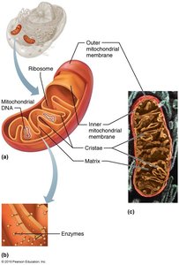
Mitochondria are known as the "power plants" of cells because they produce most of the cell’s energy molecules (ATP) via aerobic cellular respiration. They are enclosed by double membranes, with the inner membrane folded into cristae, which are embedded with proteins essential for cellular respiration. Mitochondria contain their own DNA, RNA, and ribosomes, and can divide by fission, similar to bacteria.
Function: ATP production through aerobic respiration
Structure: Outer and inner membranes, cristae, matrix
Unique Features: Own genetic material and ribosomes
Ribosomes
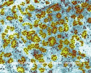
Ribosomes are nonmembranous organelles that serve as the site of protein synthesis. They are composed of protein and ribosomal RNA (rRNA) and exist in two forms:
Free ribosomes: Float freely in the cytosol; synthesize soluble proteins for use within the cell.
Membrane-bound ribosomes: Attached to the ER; synthesize proteins for incorporation into membranes, lysosomes, or for export.
Endoplasmic Reticulum (ER)
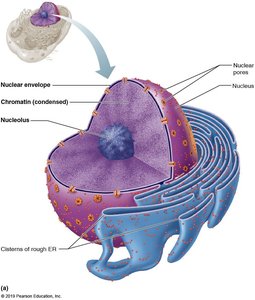
The endoplasmic reticulum (ER) is a network of interconnected membranous tubules and sacs (cisterns) continuous with the outer nuclear membrane. It exists in two forms:
Rough ER: Studded with ribosomes; site of synthesis for secreted proteins, plasma membrane proteins, and phospholipids.
Smooth ER: Lacks ribosomes; involved in lipid metabolism, detoxification, glycogen breakdown, and calcium storage.
Golgi Apparatus
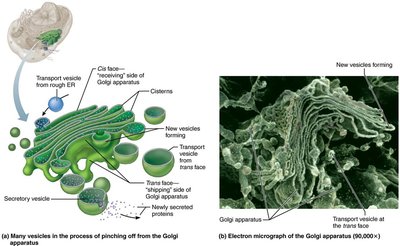
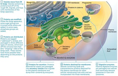
The Golgi apparatus is a stack of flattened membranous sacs that modifies, concentrates, and packages proteins and lipids from the rough ER. It directs products to their final destinations via vesicles.
Step 1: Transport vesicles from ER fuse with the cis face of the Golgi.
Step 2: Proteins/lipids are modified, tagged, sorted, and packaged.
Step 3: Final products are sent to their destinations as vesicles pinch off the trans face.
Peroxisomes
Peroxisomes are membranous sacs containing enzymes that detoxify harmful substances. They neutralize free radicals using oxidase (producing hydrogen peroxide) and catalase (converting hydrogen peroxide to water). They also participate in fatty acid metabolism.
Lysosomes
Lysosomes are spherical membranous organelles containing digestive enzymes (acid hydrolases). They digest ingested bacteria, viruses, and toxins, degrade nonfunctional organelles, and release stored substances such as glycogen and calcium. Lysosomal rupture can lead to autolysis (self-digestion).
Cytoskeleton
The cytoskeleton is an elaborate network of protein rods that provide structural support and facilitate movement of cell components. It consists of:
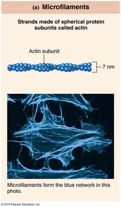
Microfilaments: Thin strands of actin; involved in cell movement and shape changes.
Intermediate filaments: Tough, insoluble protein fibers; provide mechanical strength.
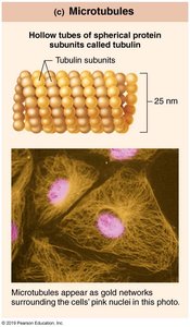
Microtubules: Hollow tubes of tubulin; determine cell shape and organelle distribution.
3.8 Cellular Extensions
Cilia and Flagella
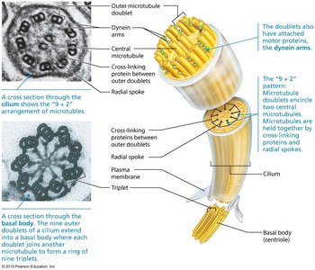
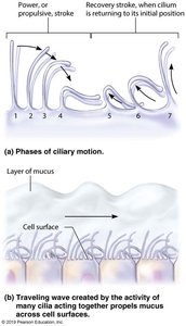
Cilia and flagella are cellular extensions that aid in movement. Cilia move substances across the cell surface, while flagella propel the cell itself (e.g., sperm cell).
Microvilli
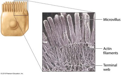
Microvilli are minute, fingerlike projections of the plasma membrane that increase surface area for absorption. They contain a core of actin filaments for structural support.
Nucleus and Genetic Material
Nucleus
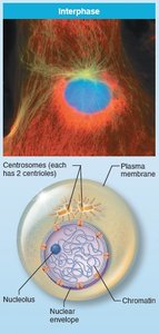
The nucleus is the largest organelle and contains the genetic library for nearly all cellular proteins. It is surrounded by a nuclear envelope and contains nucleoli and chromatin. Most cells are uninucleate, but some are multinucleate or anucleate.
Chromatin and Chromosome Structure
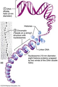
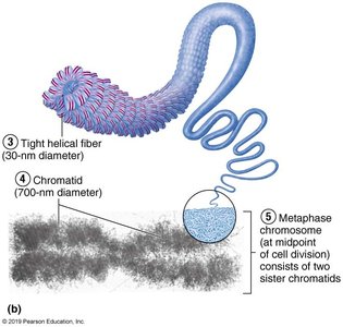
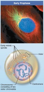
Chromatin consists of DNA wrapped around histone proteins, forming nucleosomes. During cell division, chromatin condenses into chromosomes, each consisting of two sister chromatids joined at a centromere.
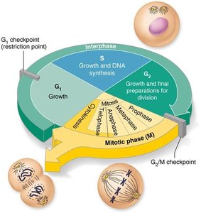
3.10 Cell Cycle
Overview of the Cell Cycle
The cell cycle is a series of events that cells go through as they grow and divide. It consists of two major periods: interphase (cell growth and DNA replication) and mitotic phase (cell division).
Interphase: Includes G1 (growth), S (DNA synthesis), and G2 (preparation for division).
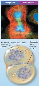
Mitotic phase (M phase): Includes mitosis (nuclear division) and cytokinesis (cytoplasmic division).
Mitosis
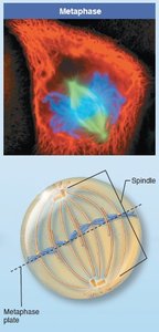
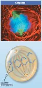
Mitosis is the process of nuclear division, ensuring each daughter cell receives a complete set of chromosomes. It consists of four stages:
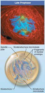
Prophase: Chromatin condenses into chromosomes; spindle apparatus forms.
Metaphase: Chromosomes align at the metaphase plate.
Anaphase: Sister chromatids separate and move toward opposite poles.
Telophase: Chromosomes decondense; nuclear envelopes reform.
Control of Cell Division
Cell division is regulated by "go" and "stop" signals, including surface-to-volume ratio, growth factors, and contact inhibition. Checkpoints (especially the G1 checkpoint) ensure proper division; failure to pass leads to G0 phase (no division).
3.11 Protein Synthesis
Genetic Code and Role of RNA
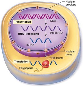
DNA contains the genetic code for protein synthesis, organized into genes. The code is a sequence of nitrogenous bases (A, G, T, C) read in triplets. RNA acts as the intermediary, with three main types:
mRNA (messenger RNA): Carries the genetic code from DNA to ribosomes.
rRNA (ribosomal RNA): Forms part of the ribosome structure.
tRNA (transfer RNA): Brings amino acids to the ribosome during translation.
Steps of Protein Synthesis
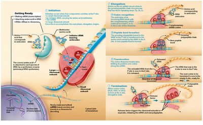
Transcription: DNA is transcribed into pre-mRNA, which is processed into mature mRNA.
Translation: mRNA is decoded by ribosomes to assemble a polypeptide chain.
3.12 Apoptosis, Autophagy, and Proteasomes
Autophagy
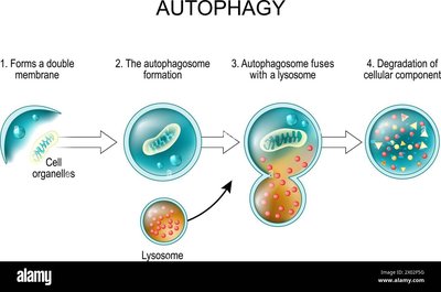
Autophagy is the process by which cells degrade and recycle nonfunctional organelles and cytoplasmic components. Autophagosomes fuse with lysosomes for degradation.
Ubiquitin-Proteasome Pathway
Proteins that are unneeded, misfolded, or damaged are tagged with ubiquitin and degraded by proteasomes, recycling amino acids and ubiquitin.
Apoptosis
Apoptosis is programmed cell death, involving activation of caspases, degradation of DNA and cytoskeleton, and phagocytosis of cell remnants. It is essential for removing damaged or unnecessary cells.
Developmental Aspects and Cell Aging
Cell Differentiation
All cells contain the same DNA, but differentiation occurs through selective gene expression, leading to specialized cell functions.
Cell Destruction and Division Rates
Cell division is necessary for growth and repair. Hyperplasia is increased cell number; atrophy is decreased cell size due to loss of stimulation or use.
Cell Aging
Cell aging theories include wear and tear, mitochondrial dysfunction, immune system decline, and genetic programming. Telomeres protect chromosome ends but shorten with each division, limiting cell lifespan. Telomerase can extend telomeres, found in germ cells and cancer cells.
