 뒤로
뒤로Epithelial Tissues: Structure, Classification, and Function
스터디 가이드 - 스마트 노트
 자료에 맞춘 맞춤형 노트, 핵심 정의, 예시, 맥락을 확장해 제공합니다.
Tissue: The Living Fabric
Introduction to Tissues
Tissues are groups of cells that are similar in structure and perform a common or related function. The study of tissues is known as histology. There are four primary tissue types in the human body:
Epithelial tissue – Covers body surfaces and lines cavities
Connective tissue – Supports, protects, and binds other tissues
Muscle tissue – Produces movement
Nervous tissue – Controls and integrates body functions
Each tissue type plays a vital role in maintaining homeostasis and the overall function of organs and organ systems.
Epithelial Tissue
Overview and Functions
Epithelial tissue forms boundaries between different environments, protects underlying structures, secretes substances, absorbs nutrients, and filters materials. It is found lining the digestive tract, covering the skin surface (epidermis), and forming glands.
Protection: Shields underlying tissues from mechanical and chemical injury.
Absorption: Uptake of nutrients and other substances (e.g., in the intestines).
Filtration: Selective permeability, especially in kidney tubules.
Secretion: Production and release of substances such as mucus, sweat, and enzymes.
Characteristics of Epithelial Tissues
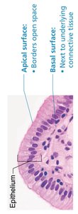
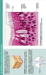
Polarity: Epithelial cells have an apical surface (exposed to the exterior or cavity) and a basal surface (attached to underlying connective tissue). The apical surface may have microvilli (to increase surface area) or cilia (for movement of substances).
Specialized Contacts: Cells are closely joined by tight junctions and desmosomes, providing structural integrity.
Supported by Connective Tissue: The basement membrane reinforces the epithelial sheet and defines its boundary.
Avascular but Innervated: Epithelial tissues lack blood vessels but are supplied by nerve fibers. Nutrients diffuse from underlying connective tissues.
Regeneration: Epithelial cells have a high regenerative capacity, rapidly replacing lost or damaged cells.
Classification of Epithelia
By Number of Cell Layers
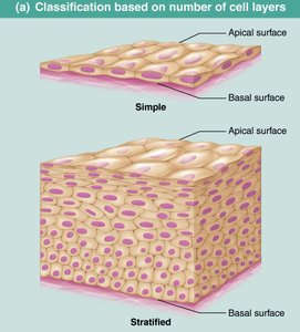
Epithelia are classified based on the number of cell layers present:
Simple epithelium: Single layer of cells; typically involved in absorption, secretion, and filtration.
Stratified epithelium: Two or more layers; primarily for protection.
By Cell Shape
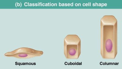
The shape of the cells at the apical surface determines the classification:
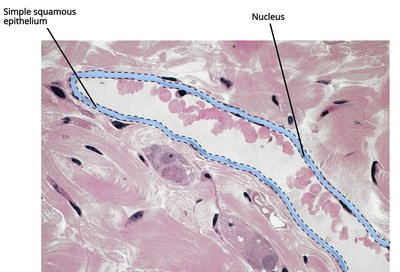
Squamous: Flattened and scale-like; nucleus is also flattened.
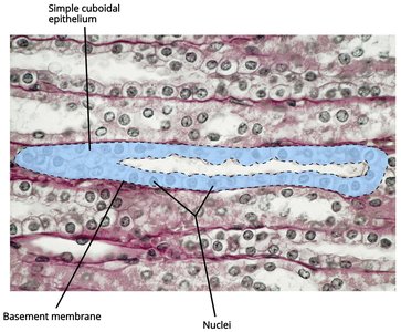
Cuboidal: Boxlike; nucleus is round.
Columnar: Tall and column-shaped; nucleus is elongated.
Types of Epithelial Tissues
Simple Epithelia
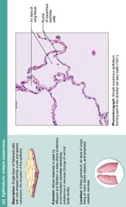
Simple Squamous Epithelium: Single layer of flat cells; allows for rapid diffusion and filtration. Found in air sacs of lungs, lining of heart, blood vessels, and lymphatic vessels.
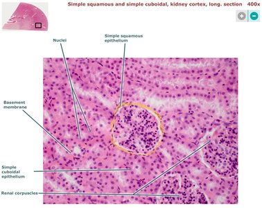
Simple Cuboidal Epithelium: Single layer of cube-shaped cells; functions in secretion and absorption. Common in kidney tubules and ducts of small glands.

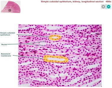
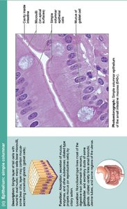
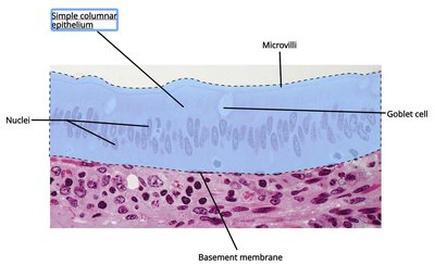
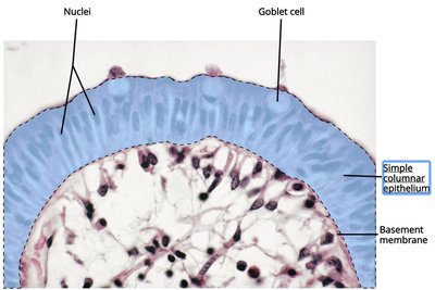
Simple Columnar Epithelium: Single layer of tall, column-like cells; involved in absorption and secretion. May have microvilli (for absorption) and goblet cells (secreting mucus). Found in the digestive tract, gallbladder, and some ducts.
Stratified Epithelia
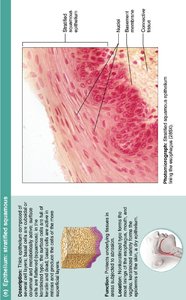
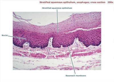
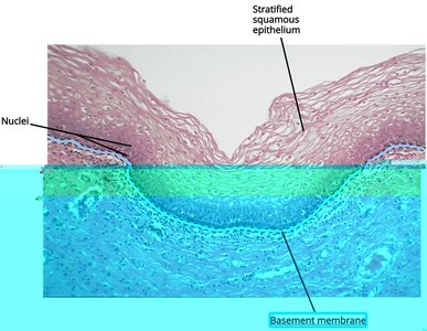
Stratified Squamous Epithelium: Multiple layers of cells with squamous cells at the apical surface; protects underlying tissues in areas subject to abrasion. Found in the esophagus, mouth, vagina, and skin (keratinized type).
Summary Table: Epithelial Tissue Types
Type | Structure | Function | Location |
|---|---|---|---|
Simple Squamous | Single layer, flat cells | Diffusion, filtration | Air sacs of lungs, lining of heart, blood vessels |
Simple Cuboidal | Single layer, cube-shaped cells | Secretion, absorption | Kidney tubules, ducts of small glands |
Simple Columnar | Single layer, tall cells | Absorption, secretion of mucus/enzymes | Digestive tract, gallbladder |
Stratified Squamous | Multiple layers, apical cells squamous | Protection | Esophagus, mouth, vagina, skin |
Key Terms
Apical surface: The free surface exposed to the body exterior or the cavity of an internal organ.
Basal surface: The lower, attached surface facing the underlying connective tissue.
Basement membrane: A thin, fibrous, extracellular matrix that separates the epithelium from underlying connective tissue.
Microvilli: Fingerlike extensions of the plasma membrane that increase surface area for absorption.
Cilia: Hairlike projections that move substances along the surface of the cell.
Goblet cells: Unicellular glands that secrete mucus, commonly found in columnar epithelium.
