뒤로
뒤로Equilibrium and Hearing: Anatomy & Physiology Study Notes
스터디 가이드 - 스마트 노트
 자료에 맞춘 맞춤형 노트, 핵심 정의, 예시, 맥락을 확장해 제공합니다.
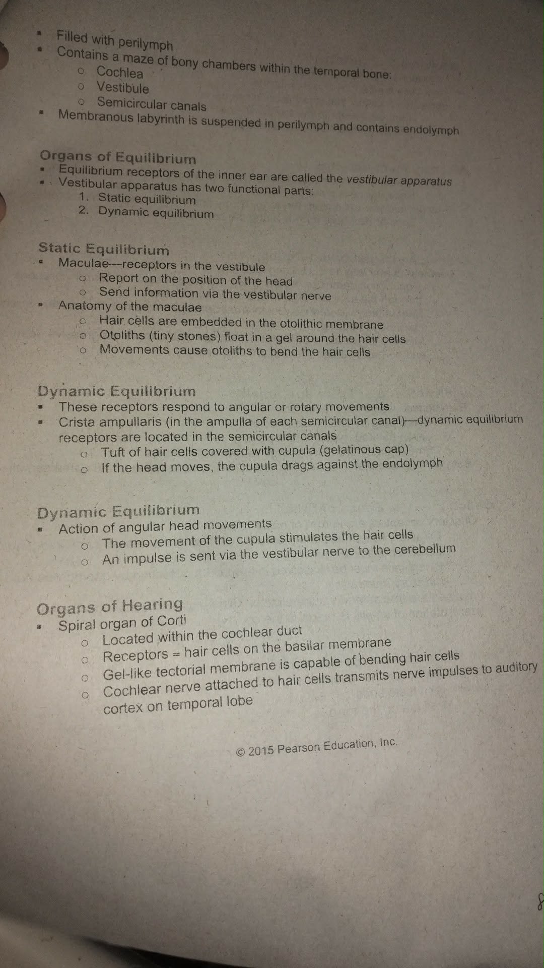
Organs of Equilibrium
Vestibular Apparatus
The vestibular apparatus is a key structure in the inner ear responsible for maintaining balance and equilibrium. It consists of two main functional parts: static equilibrium and dynamic equilibrium.
Static Equilibrium: Detects the position of the head relative to gravity.
Dynamic Equilibrium: Detects rotational or angular movements of the head.
Static Equilibrium
Static equilibrium is managed by the maculae, which are receptors located in the vestibule of the inner ear. These structures report the position of the head and send information via the vestibular nerve.
Maculae Anatomy:
Hair cells are embedded in the otolithic membrane.
Otoliths (tiny stones) float in a gel around the hair cells.
Movements cause otoliths to bend the hair cells, triggering nerve impulses.
Dynamic Equilibrium
Dynamic equilibrium receptors respond to angular or rotary movements. The crista ampullaris is located in the ampulla of each semicircular canal and is responsible for detecting these movements.
Crista Ampullaris Anatomy:
Tuft of hair cells covered with a cupula (gelatinous cap).
If the head moves, the cupula drags against the endolymph, stimulating the hair cells.
An impulse is sent via the vestibular nerve to the cerebellum for processing.
Organs of Hearing
Spiral Organ of Corti
The spiral organ of Corti is the primary hearing organ located within the cochlear duct. It contains specialized hair cells that detect sound vibrations.
Receptors: Hair cells on the basilar membrane.
Tectorial Membrane: Gel-like membrane capable of bending hair cells.
Cochlear Nerve: Attached to hair cells, transmits nerve impulses to the auditory cortex in the temporal lobe.
Anatomy of the Inner Ear
Key Structures
The inner ear is filled with perilymph and contains a maze of bony chambers within the temporal bone:
Cochlea: Responsible for hearing.
Vestibule: Responsible for static equilibrium.
Semicircular Canals: Responsible for dynamic equilibrium.
The membranous labyrinth is suspended in perilymph and contains endolymph, which is essential for the function of equilibrium and hearing receptors.
Summary Table: Inner Ear Structures and Functions
Structure | Main Function | Key Feature |
|---|---|---|
Cochlea | Hearing | Contains organ of Corti |
Vestibule | Static Equilibrium | Contains maculae |
Semicircular Canals | Dynamic Equilibrium | Contains crista ampullaris |
Example: How Equilibrium Works
When you tilt your head, otoliths move and bend the hair cells in the maculae, sending signals to your brain about head position.
When you spin or rotate your head, the cupula in the semicircular canals moves, stimulating hair cells and sending information about movement to the cerebellum.
Additional info:
The cerebellum is the brain region responsible for processing equilibrium information and coordinating balance.
Damage to any of these structures can result in dizziness, loss of balance, or hearing impairment.