 뒤로
뒤로Foundations: An Introduction to Anatomy – Study Notes
스터디 가이드 - 스마트 노트
 자료에 맞춘 맞춤형 노트, 핵심 정의, 예시, 맥락을 확장해 제공합니다.
Foundations: An Introduction to Anatomy
Introduction to Anatomy and Physiology
Anatomy is the study of the structure of the human body, including both external and internal features, and the relationships between body parts. Physiology is the study of how the body functions, focusing on the mechanisms that allow anatomical structures to perform their roles. Understanding anatomy provides essential clues to physiological functions, as structure and function are closely related.
Microscopic Anatomy
Microscopic anatomy examines structures that cannot be seen without magnification. It includes:
Cytology: The study of the internal structure of individual cells, the smallest units of life.
Histology: The study of tissues, which are groups of specialized cells working together to perform specific functions.
Microscopic anatomy is foundational to understanding how cells form tissues, which in turn form organs.
Gross Anatomy (Macroscopic Anatomy)
Gross anatomy involves the study of structures visible to the unaided eye. It can be approached in several ways:
Surface Anatomy: Study of general anatomical form and superficial markings.
Regional Anatomy: Study of all structures in a specific area (e.g., head, neck, trunk).
Systemic Anatomy: Study of the organ systems of the body (e.g., cardiovascular, digestive).
Other Types of Anatomical Studies
Developmental Anatomy: Examines structural changes over time, including embryology (development from conception through the first eight weeks).
Comparative Anatomy: Considers anatomical similarities and differences among different animal species.
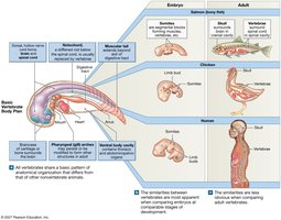
Clinical Anatomy: Studies anatomical changes during pathological illnesses.
Surgical Anatomy: Focuses on anatomical landmarks important for surgical procedures.
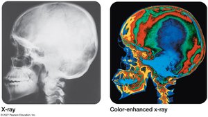
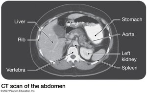
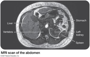
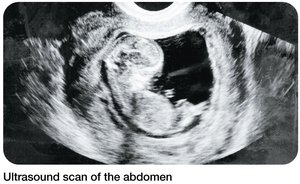
Radiographic Anatomy: Uses imaging techniques (e.g., x-rays, CT, MRI, ultrasound) to study anatomy.
Cross-sectional Anatomy: Uses radiographic techniques to study cross sections of the body.
Levels of Organization in the Human Body
Overview of Levels
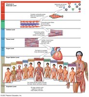
The human body is organized into a hierarchy of structural levels, from the simplest to the most complex:
Chemical/Molecular Level: Atoms combine to form molecules, which are the building blocks of cells.
Cellular Level: Cells are the smallest living units, containing organelles that perform specific functions.
Tissue Level: Tissues are groups of similar cells working together (e.g., epithelial, muscular, neural, connective).
Organ Level: Organs are combinations of tissues performing specific functions (e.g., the heart).
Organ System Level: Organ systems are groups of organs working together (e.g., digestive, cardiovascular).
Organism Level: The human organism is composed of all organ systems working together to maintain life.
Chemical/Molecular Level
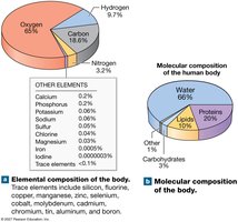
Over a dozen elements are present in the body, but four (hydrogen, oxygen, carbon, nitrogen) make up 99% of the body. Major classes of compounds include:
Water: 66%
Proteins: 20%
Lipids: 10%
Carbohydrates: 3%
Cellular, Tissue, and Organ Levels
Cells are the basic living units, containing organelles. Tissues are made of similar cells, and organs are combinations of tissues. For example, the heart contains epithelial, muscular, neural, and connective tissues.
Organ System and Organism Levels
Organ systems are combinations of organs with related functions. For example, the digestive system includes the stomach, intestines, liver, gallbladder, and pancreas. The cardiovascular system includes the heart and blood vessels. Humans have 11 organ systems that work together to maintain homeostasis and life.
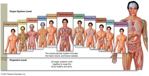
Introduction to Organ Systems
Key Characteristics of Living Organisms
Responsiveness: Ability to respond to internal and external changes, including adaptability (long-term adjustments).
Growth and Differentiation: Increase in cell size and number; cells become specialized for specific functions.
Reproduction: Production of new generations of the same organism.
Metabolism and Excretion: All chemical reactions in the body, including anabolism (building molecules), catabolism (breaking down molecules), absorption, respiration, and excretion (removal of waste).
Movement: Internal (e.g., movement of food or blood) and external (e.g., walking).
The Language of Anatomy
Importance of Anatomical Terminology
Precise vocabulary, often derived from Latin and Greek, is essential for clear communication in anatomy. Terms describe positions, directions, and regions of the body.
Superficial Anatomy: Anatomical Landmarks and Position
The anatomical position is the standard reference: standing, feet flat, hands at sides, palms facing forward. All anatomical descriptions refer to this position. Supine means lying face up; prone means lying face down.
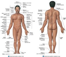
Superficial Anatomy: Anatomical Regions
The body is divided into major regions for descriptive purposes, such as head, neck, trunk, upper limb, and lower limb.
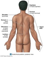
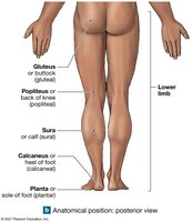
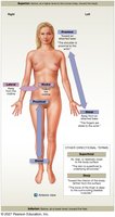
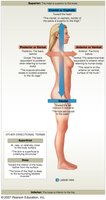
Superficial Anatomy: Directional Terms
Common directional terms include:
Superior (above), Inferior (below)
Anterior (ventral) (front), Posterior (dorsal) (back)
Medial (toward midline), Lateral (away from midline)
Superficial (toward surface), Deep (away from surface)
Proximal (closer to trunk), Distal (farther from trunk)
Superficial Anatomy: Planes and Sections
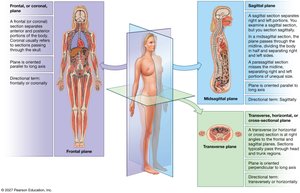
Sectional planes are used to describe cuts through the body:
Sagittal plane: Divides body into left and right (midsagittal = equal halves, parasagittal = unequal).
Transverse plane: Divides body into superior and inferior (horizontal sections).
Frontal (coronal) plane: Divides body into anterior and posterior sections.
Serial Reconstruction
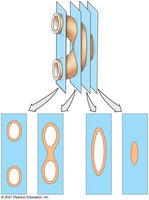
Serial reconstruction involves making a series of sections at small intervals to visualize three-dimensional structures.
Sectional Anatomy: Body Cavities
Major Body Cavities
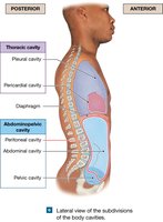
Body cavities are internal chambers that protect organs and allow them to change shape. The two major cavities are:
Posterior (dorsal) cavity: Includes cranial and spinal cavities.
Anterior (ventral) cavity: Includes thoracic and abdominopelvic cavities, separated by the diaphragm.
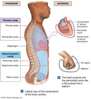
Thoracic cavity: Contains heart, lungs, esophagus, trachea; subdivided into pleural and pericardial cavities.
Abdominopelvic cavity: Contains abdominal and pelvic organs; lined by the peritoneum.
Serous Membranes of Body Cavities
Serous membranes line body cavities and cover organs, reducing friction:
Pleural cavity: Visceral and parietal pleura surround the lungs.
Pericardial cavity: Visceral and parietal layers of the serous pericardium surround the heart.
Peritoneal cavity: Visceral and parietal peritoneum surround abdominal organs.
Additional info: Understanding the organization of the human body and the terminology used in anatomy is essential for further study in all areas of health and medicine. Mastery of these foundational concepts enables clear communication and accurate identification of structures and their functions.
