뒤로
뒤로Histology and Tissue Organization: Foundations of Anatomy & Physiology
스터디 가이드 - 스마트 노트
 자료에 맞춘 맞춤형 노트, 핵심 정의, 예시, 맥락을 확장해 제공합니다.
Histology and Levels of Organization
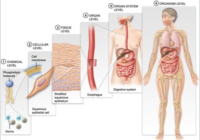
Overview of Levels of Organization
Understanding the human body begins with recognizing its hierarchical structure, from the smallest chemical components to the complete organism. Each level builds upon the previous, contributing to the complexity and function of living systems.
Chemical Level: Atoms and molecules form the building blocks of cells.
Cellular Level: Cells are the basic units of life, each with specialized functions.
Tissue Level: Groups of similar cells and extracellular matrix (ECM) form tissues with specific roles.
Organ Level: Different tissues combine to form organs, each with distinct functions.
Organ System Level: Organs work together in systems (e.g., digestive system) to perform complex tasks.
Organism Level: All organ systems function together to sustain the living human body.
Cell Junctions and Tissue Integrity
Types of Cell Junctions
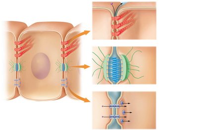
Cell junctions are specialized structures that connect adjacent cells, providing structural integrity and facilitating communication within tissues.
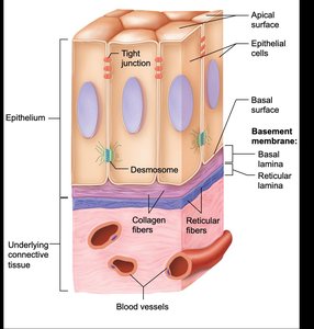
Tight Junctions: Seal spaces between cells, making tissues impermeable to most substances.
Desmosomes: Anchor cells together, increasing tissue resistance to mechanical stress.
Gap Junctions: Allow ions and small molecules to pass directly between cells, enabling rapid communication.
Extracellular Matrix (ECM)
Components and Functions of ECM
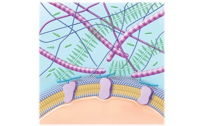
The extracellular matrix is a complex network of proteins and carbohydrates that provides structural and biochemical support to surrounding cells.
Ground Substance: Contains glycosaminoglycans (GAGs), proteoglycans, and glycoproteins, which help retain water and resist compression.
Protein Fibers: Includes collagen (strength), elastic fibers (flexibility), and reticular fibers (supportive meshwork).
Classification of Tissues
The Four Basic Tissue Types
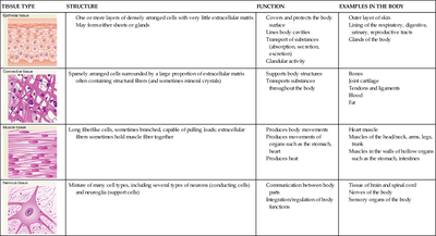
Tissues are classified based on their structure and function. The four primary tissue types are epithelial, connective, muscle, and nervous tissue.
Tissue Type | Structure | Function | Examples in the Body |
|---|---|---|---|
Epithelial Tissue | One or more layers of densely arranged cells with very little extracellular matrix; may form sheets or glands | Covers and protects the body, lines body cavities, transports substances, forms glands | Outer layer of skin, lining of respiratory, digestive, urinary, and reproductive tracts, glands |
Connective Tissue | Sparsely arranged cells surrounded by a large proportion of extracellular matrix; often containing structural fibers | Supports body structures, transports substances, connects tissues | Bones, joint cartilage, tendons, ligaments, fat |
Muscle Tissue | Long, fiberlike cells, sometimes branched, capable of pulling loads; extracellular fibers hold muscle fibers together | Produces body movements, produces heat | Heart muscle, muscles of the head, neck, arms, legs, trunk, walls of hollow organs |
Nervous Tissue | Mixture of many cell types, including neurons and neuroglia (supporting cells) | Communication between body parts, regulation of body functions | Brain, spinal cord, nerves |
Epithelial Tissue Structure
Organization and Features
Epithelial tissue forms protective barriers and is characterized by closely packed cells with minimal ECM. It has distinct surfaces and is supported by a basement membrane.
Apical Surface: Exposed to the body exterior or cavity.
Basal Surface: Attached to the underlying connective tissue via the basement membrane.
Basement Membrane: Composed of basal lamina and reticular lamina, providing support and filtration.
Glandular Epithelium
Unicellular and Multicellular Glands
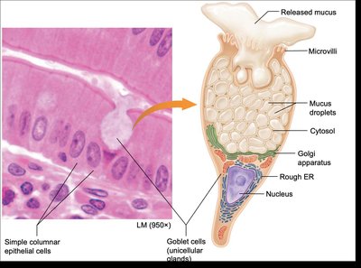
Glandular epithelium specializes in secretion. Glands can be unicellular (e.g., goblet cells) or multicellular (e.g., sweat glands).
Goblet Cells: Unicellular glands that secrete mucus, found in the lining of the respiratory and digestive tracts.
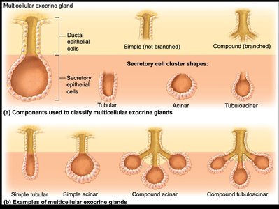
Multicellular Glands: Composed of multiple cells forming ducts and secretory units; classified by duct structure (simple or compound) and shape (tubular, acinar, tubuloacinar).
Modes of Secretion in Exocrine Glands
Exocrine glands release their products by different mechanisms:
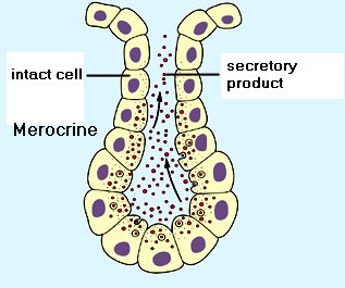
Merocrine Secretion: Secretory products are released by exocytosis; the cell remains intact (e.g., sweat glands).
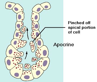
Apocrine Secretion: The apical portion of the cell pinches off, releasing cytoplasm along with the secretion (e.g., mammary glands).
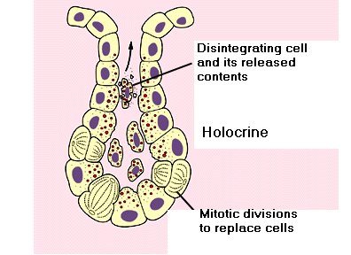
Holocrine Secretion: The entire cell disintegrates to release its contents (e.g., sebaceous glands).
Tissues in Organs: Example of the Trachea
Integration of Multiple Tissue Types
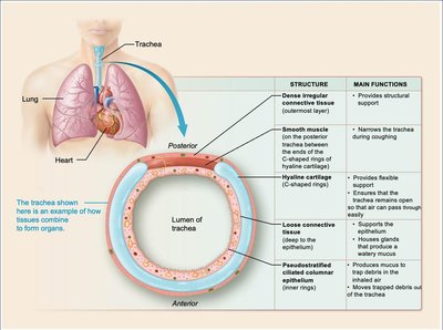
Organs are composed of multiple tissue types working together to perform specific functions. The trachea, for example, contains connective, muscle, cartilage, and epithelial tissues, each contributing to its structure and function.
Dense Irregular Connective Tissue: Provides structural support.
Smooth Muscle: Narrows the trachea during coughing.
Hyaline Cartilage: Maintains an open airway.
Loose Connective Tissue: Supports the epithelium and blood vessels.
Pseudostratified Ciliated Columnar Epithelium: Moves mucus and debris out of the trachea.
Membranes and Tissue Repair
Types of Membranes
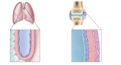
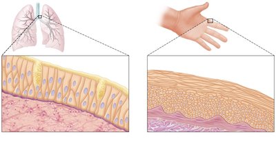
Membranes are thin layers of tissue that cover surfaces, line cavities, and divide spaces or organs. Major types include serous, synovial, mucous, and cutaneous membranes.
Serous Membranes: Line body cavities not open to the exterior; secrete serous fluid for lubrication.
Synovial Membranes: Line joint cavities; secrete synovial fluid for joint lubrication.
Mucous Membranes: Line body cavities open to the exterior; secrete mucus for protection and moisture.
Cutaneous Membrane: The skin; protects the body surface.
Tissue Repair Mechanisms
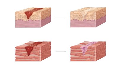
Tissues repair themselves by regeneration (replacement with the same cell type) or fibrosis (replacement with scar tissue).
Regeneration: Restores normal function; common in epithelial tissues.
Fibrosis: Forms scar tissue; common in tissues with limited regenerative capacity (e.g., cardiac muscle).
Summary Table: Epithelial Tissue Types
Classification by Layers and Cell Shape
Epithelial tissues are classified based on the number of cell layers and the shape of the cells at the apical surface.
Simple Epithelium: Single layer of cells; allows for absorption, secretion, and filtration.
Stratified Epithelium: Multiple layers; provides protection against abrasion.
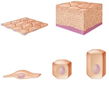
Cell Shapes: Squamous (flat), cuboidal (cube-shaped), columnar (tall and column-like).