 뒤로
뒤로Histology and Tissue Types: Structure, Function, and Identification
스터디 가이드 - 스마트 노트
 자료에 맞춘 맞춤형 노트, 핵심 정의, 예시, 맥락을 확장해 제공합니다.
Histology and Tissues
Introduction to Tissues
Tissues are groups of cells working together to perform a common function and serve as the fundamental building blocks of organs. Each tissue consists of cells and an extracellular matrix (ECM), which provides structural and functional support. The study of tissues, known as histology, is essential for understanding organ structure and function.
Tissue Definition: Group of cells + extracellular matrix (ECM)
Functions: Vary by tissue type (support, movement, communication, etc.)
Classification of Tissue Types
There are four primary tissue types in the human body, each with distinct functions and locations:
Epithelial Tissue: Covering and lining surfaces
Nervous Tissue: Communication and control
Connective Tissue: Support, transport, energy storage, etc.
Muscle Tissue: Movement
Epithelial Tissues
General Characteristics
Epithelial tissues cover body surfaces and line internal cavities. They are tightly packed with minimal ECM, have high regenerative capacity, and possess distinct apical (top/exposed) and basal (bottom/attached) surfaces.
Functions: Protection, absorption, secretion, filtration
Regeneration: Rapid cell turnover
Naming Epithelial Tissues
Epithelial tissues are named based on the number of cell layers and cell shape:
Cell Layers:
Simple: Single layer
Stratified: Multiple layers
Cell Shape:
Squamous: Flattened
Cuboidal: Cube-shaped
Columnar: Tall, rectangular
Simple Squamous Epithelium
Single layer of flattened cells specialized for filtration and diffusion. Found in locations such as alveoli of lungs and lining of blood vessels.
Simple Cuboidal Epithelium
Single layer of cube-shaped cells, often arranged in rings. Functions in secretion and absorption. Commonly found in kidney tubules and glandular ducts.
Simple Columnar Epithelium
Single layer of column-shaped cells, specialized for secretion and absorption. Found in the lining of the digestive tract (stomach, intestines).
Example: Intestinal lining for nutrient absorption
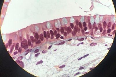
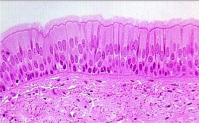
Pseudostratified Ciliated Columnar Epithelium
Appears stratified but is a single layer of columnar cells with cilia on the apical surface. Functions in protection, secretion, and movement of mucus. Located in the respiratory tract (trachea, bronchi).
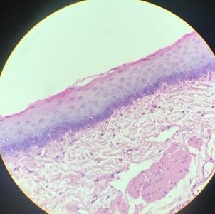
Stratified Squamous Epithelium
Multiple layers of flattened cells providing protection against abrasion and friction. Found in the skin, mouth, esophagus, and vagina.
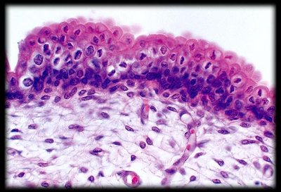
Transitional Epithelium
Multiple layers of polyhedral cells capable of stretching and recoiling. Specialized for flexibility, found in the urinary bladder and ureters.
Nervous Tissue
Structure and Function
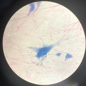
Nervous tissue is responsible for communication and control throughout the body. It consists of two main cell types:
Neurons: Large, often branched cells that receive and transmit electrical signals
Neuroglia: Small support cells with varied functions (protection, insulation, support)
Located in the brain, spinal cord, and peripheral nerves.
Connective Tissues
General Characteristics
Connective tissues consist of cells surrounded by abundant extracellular matrix (fibers and ground substance). They serve various functions and are subdivided into proper, dense, supportive, and fluid connective tissues.
Functions: Support, protection, transport, energy storage
Proper Connective Tissues
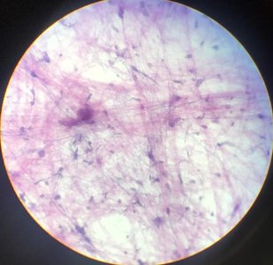
Areolar Connective Tissue
Contains fibers and fibroblasts with abundant ground substance. Provides support and cushioning, found beneath epithelial layers and around organs.
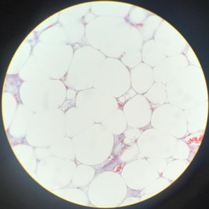
Adipose Connective Tissue
Loosely packed adipocytes that store triglycerides. Functions in long-term energy storage and protection. Located under the skin and around organs.
Dense Connective Tissues
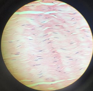
Dense Regular Connective Tissue
Parallel arrangement of collagen fibers with sporadic fibroblasts. Resists tension in a single direction. Found in tendons and ligaments.
Dense Irregular Connective Tissue
Collagen fibers not arranged in parallel, with fibroblasts. Resists tension in multiple directions. Located in dermis of skin and organ capsules.
Supportive Connective Tissues
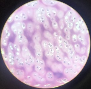
Hyaline Cartilage
Glassy matrix containing chondrocytes in lacunae. Provides support and is found in joints, nose, and trachea.
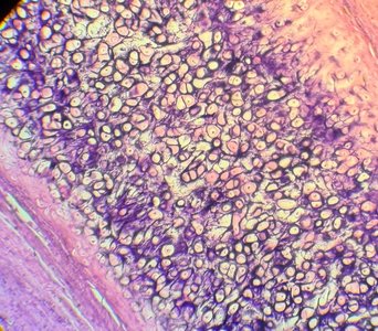
Elastic Cartilage
Chondrocytes in lacunae housed in a dense network of elastic fibers. Provides strength and flexibility. Located in the external ear and epiglottis.
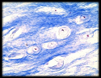
Fibrocartilage
Fibrous matrix containing chondrocytes in lacunae. Resists compression. Found in intervertebral discs and pubic symphysis.
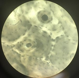
Compact Bone (Osseous Connective Tissue)
Adjacent cylindrical units (osteons) containing osteocytes in lacunae. Functions in support, movement, and protection. Located in bones throughout the body.
Fluid Connective Tissue
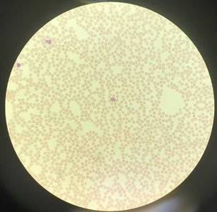
Blood
Formed elements suspended in a liquid matrix (plasma). Functions in transport of materials and protection (immune and hemostasis). Found in blood vessels throughout the body.
Muscle Tissues
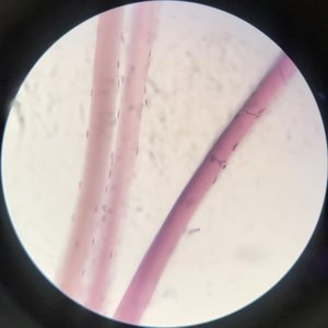
Skeletal Muscle Tissue
Long, cylindrical, striated, unbranched fibers with multiple nuclei. Voluntarily controlled. Functions in voluntary movement, posture, and ventilation/breathing. Located in muscles attached to bones.
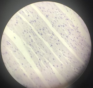
Cardiac Muscle Tissue
Short, branched, cylindrical, striated fibers connected by intercalated discs. Involuntarily controlled. Functions to pump blood through the cardiovascular system. Located in the heart.
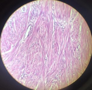
Smooth Muscle Tissue
Spindle-shaped cells, non-striated, involuntarily controlled. Functions in movement of materials and regulation of organ diameter. Found in walls of hollow organs (intestines, blood vessels).
Summary Table: Tissue Types and Functions
Tissue Type | Main Function | Location Example |
|---|---|---|
Epithelial | Protection, absorption, secretion | Skin, lining of GI tract |
Connective | Support, transport, energy storage | Bone, blood, fat |
Muscle | Movement | Skeletal muscles, heart, intestines |
Nervous | Communication, control | Brain, spinal cord, nerves |
Additional info: Tissue identification in histology relies on recognizing cell shape, arrangement, and matrix characteristics under the microscope. Understanding these features is essential for clinical and laboratory applications.
