 뒤로
뒤로Histology and Tissues: The Living Fabric – Study Notes
스터디 가이드 - 스마트 노트
 자료에 맞춘 맞춤형 노트, 핵심 정의, 예시, 맥락을 확장해 제공합니다.
Histology and Tissues: The Living Fabric
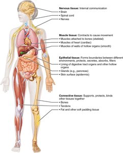
Overview of Tissue Types
Tissues are groups of cells similar in structure and function, forming the fabric of the body. There are four primary tissue types in the human body: epithelial, connective, muscle, and nervous tissue. Each type has distinct roles essential for maintaining homeostasis and supporting life.
Epithelial Tissue: Covers surfaces, lines cavities, and forms glands.
Connective Tissue: Supports, protects, and binds other tissues.
Muscle Tissue: Contracts to produce movement.
Nervous Tissue: Detects stimuli and transmits electrical signals.
Epithelial Tissue
General Characteristics
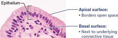
Epithelial tissue forms boundaries between different environments, covering surfaces and lining cavities. It is organized into two main forms: covering/lining epithelium and glandular epithelium.
Tightly packed cells ensure barrier and absorptive/secretory functions, maintained by tight junctions and desmosomes.
Polarity: Cells have apical (free) and basolateral (attached) surfaces with distinct functions.
Highly regenerative: Rapidly replaces damaged cells; avascular but innervated.
Classification of Epithelial Tissue
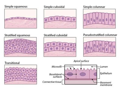
Epithelia are classified by the number of cell layers and the shape of cells at the apical surface:
Simple: Single layer of cells
Stratified: Two or more layers
Pseudostratified: Appears multilayered but is a single layer
Squamous: Flat, scale-like
Cuboidal: Box-like
Columnar: Tall, rectangular
Types of Epithelia
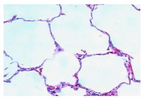
Simple Squamous Epithelium
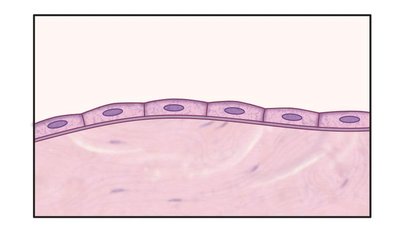
Composed of a single layer of flat cells with flattened nuclei, this tissue is specialized for filtration and diffusion.
Location: Air sacs of lungs, glomeruli, blood vessel and capillary lumens, body cavity linings.
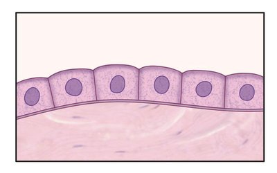
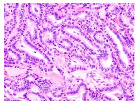
Simple Cuboidal Epithelium
Consists of a single layer of cube-shaped cells with large, round nuclei. Functions in secretion and absorption.
Location: Ducts and glands, kidney tubules, ovary, thyroid.
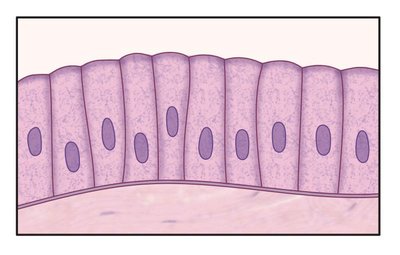
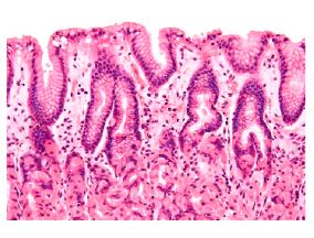
Simple Columnar Epithelium
Composed of a single layer of tall, rectangular cells with elongated nuclei. Microvilli (for absorption) and cilia (for propulsion) are common on the apical surface. Functions in absorption and secretion.
Location: Digestive tract lining, respiratory tract lining.
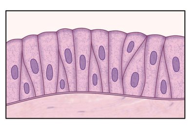
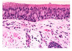
Pseudostratified Columnar Epithelium
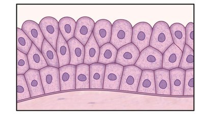
Appears to have multiple layers due to nuclei at different heights, but all cells rest on the basement membrane. Functions in secretion and propulsion (often ciliated).
Location: Male sperm duct, respiratory tract, large glands.
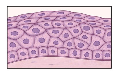
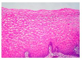
Stratified Squamous Epithelium
Multiple layers of squamous cells; surface cells are flat and keratinized, basal cells are cuboidal and mitotically active. Provides protection against abrasion.
Location: Skin, lining of esophagus, vagina, mouth.
Stratified Cuboidal/Columnar Epithelium
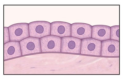
Two or more layers of cuboidal or columnar cells, functioning mainly in secretion.
Location: Male sperm duct, sweat and mammary glands, pharynx, male urethra.
Transitional Epithelium
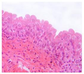
Has characteristics of both stratified squamous and cuboidal epithelia. Surface cells are rounded and can stretch, allowing for storage of urine.
Location: Lining of urinary tract (ureter, bladder, urethra).
Glandular Epithelium
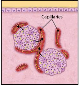
Glands are classified as endocrine (secrete hormones into capillaries) or exocrine (secrete products into ducts).
Endocrine glands: Ductless, secrete hormones directly into the bloodstream.
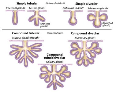
Exocrine glands: Secrete products onto body surfaces or into body cavities via ducts.
Functional Classification of Exocrine Glands
Type | Mechanism | Example |
|---|---|---|
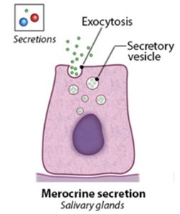
Merocrine | Secretion by exocytosis | Salivary glands |
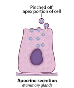
Apocrine | Secretion by pinching off part of cell | Mammary glands |
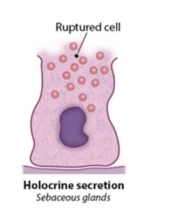
Holocrine | Secretion by cell rupture | Sebaceous glands |
Connective Tissue
General Characteristics and Functions
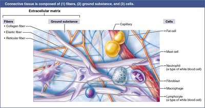
Connective tissue is the most abundant and widely distributed tissue type. It supports, protects, and binds other tissues, and is classified into four main types: connective tissue proper, cartilage, bone, and blood.
Protection: Shields tissues and organs.
Structural support: Provides framework for the body.
Energy storage and insulation: Adipose tissue stores fat.
Transport: Blood transports nutrients and gases.
Cells and Fibers of Connective Tissue
Cells: Fibroblasts/cytes (connective tissue proper), chondroblasts/cytes (cartilage), osteoblasts/cytes (bone), adipocytes (fat), blood cells (immunity).
Fibers: Collagen (strength), elastin (flexibility), reticulin (supportive meshwork).
Types of Loose Connective Tissue
Areolar Tissue
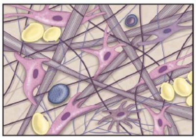
Loosely packed with all fiber types, fibroblasts, and immune cells. Cushions organs and supports immune defense.
Location: Under epithelial layers, around organs.
Adipose Tissue
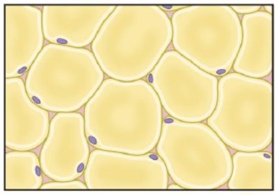
Matrix crowded by adipocytes, serving as insulation, protection, and energy storage.
Location: Around major organs, under the skin (subcutaneous layer).
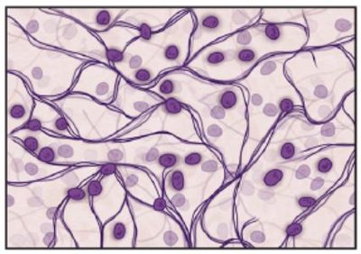
Reticular Tissue
Meshwork of reticular fibers supporting immune cells and tissues.
Location: Lymph nodes, bone marrow, spleen.
Types of Dense Connective Tissue
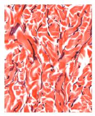
Dense Regular Connective Tissue
Fibroblasts in a regularly ordered assembly of collagen fibers, resisting pulling stress.
Location: Tendons (muscle to bone), ligaments (bone to bone).
Dense Irregular Connective Tissue
Irregularly arranged collagen fibers with fibroblasts, resisting force in multiple directions.
Location: Dermis, joint capsules, digestive tract linings.
Elastic Connective Tissue
Dense regular tissue enriched with elastin fibers, allowing stretch and recoil.
Location: Walls of arteries, bronchial tubes, some ligaments.
Types of Cartilage
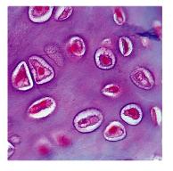
Hyaline Cartilage
Flexible, firm gel matrix secreted by chondroblasts, later embedded as chondrocytes. Cushions and supports tissues and organs.
Location: Nose, trachea, ribs, ends of long bones, embryonic skeleton.
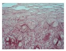
Elastic Cartilage
Similar to hyaline but with more elastin fibers, providing flexibility and stretch.
Location: Outer ear, epiglottis.
Fibrocartilage
Hyaline matrix with more organized collagen fibers, resisting compressive force.
Location: Intervertebral discs, menisci, pubic symphysis.
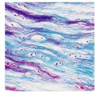
Specialized Connective Tissues
Bone
Calcified collagen matrix with embedded osteocytes, highly vascular, functions in calcium storage and muscle attachment. Spongy bone contains marrow for blood formation.
Location: Skeleton.
Blood
Composed of erythrocytes, leukocytes, and platelets in a plasma matrix. Main function is transport of gases, nutrients, and waste.
Location: Arteries, veins, capillaries.
Muscle Tissue
General Characteristics
Muscle tissue produces movement, maintains posture, and generates heat. Muscle cells (fibers) require large amounts of ATP. There are three types: skeletal, cardiac, and smooth muscle.
Skeletal muscle: Voluntary movement, attached to bones, striated, multinucleated.
Cardiac muscle: Involuntary, heart wall, striated, branched, intercalated discs.
Smooth muscle: Involuntary, walls of hollow organs, non-striated, spindle-shaped cells.
Nervous Tissue
General Characteristics
Nervous tissue is specialized for communication and control, detecting stimuli and transmitting electrical signals. It is composed of neurons and glial cells.
Neurons: Receive sensory input, transmit motor output, form synapses.
Glial cells: Support, protect, and nourish neurons.
Body Membranes
Types of Membranes
Body membranes are multicellular sheets composed of epithelial and connective tissue, forming simple organs. There are three main types:
Cutaneous membrane: Skin; dry, exposed to air.
Mucous membrane: Lines cavities open to the exterior (digestive, respiratory, urogenital); wet, adapted for absorption and secretion.
Serous membrane: Lines closed ventral body cavities; allows organs to slide (pleura, pericardium, peritoneum).
Tissue Repair
Phases of Tissue Repair
Tissue repair restores homeostasis and prevents infection. The process involves three main steps:
Inflammation: White blood cells and clotting factors enter the damage site, forming a clot (scab).
Organization (Restoration of blood supply): Fibroblasts lay down temporary extracellular matrix, new capillaries grow, and epithelium forms a new tissue layer.
Regeneration and Fibrosis: Scab falls off; scar tissue may remain depending on severity.
Tissue Healing Capacity
Different tissues heal at different rates:
Heals well: Epithelial tissue, bone, areolar connective tissue, dense irregular connective tissue, blood-forming tissue.
Moderate: Smooth muscle, dense regular connective tissue.
Weak: Skeletal muscle, cartilage.
Poor/None: Cardiac muscle, nervous tissue in brain and spinal cord.
Role of Stem Cells
Stem cells provide tissues with regenerative capacity by self-renewing and differentiating into mature cell types. Intermediate populations increase the number of mature cells produced.
Developmental Aspects of Tissue
Embryonic Development
During embryonic development, three primary germ layers (ectoderm, mesoderm, endoderm) specialize to form the primary tissues that make up all body organs.
Ectoderm: Forms nervous tissue and epidermis.
Mesoderm: Forms muscle, connective tissue, and some epithelial tissues.
Endoderm: Forms epithelial linings of digestive and respiratory tracts.
References: Marieb EN, Hoehn K. Human Anatomy & Physiology. 12th ed. Pearson; 2025. Redden J, Crivello J. Anatomy & Physiology in Context. 1st ed. Top Hat; 2024.
