 뒤로
뒤로Histology and Tissues: The Living Fabric – Study Notes for Anatomy & Physiology
스터디 가이드 - 스마트 노트
 자료에 맞춘 맞춤형 노트, 핵심 정의, 예시, 맥락을 확장해 제공합니다.
Histology and Tissues: The Living Fabric
Overview of Tissue Types
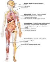
Tissues are groups of cells similar in structure and function, forming the fabric of the human body. There are four primary tissue types, each with distinct roles in maintaining body function and structure.
Epithelial Tissue: Covers surfaces and lines cavities; forms boundaries between environments.
Connective Tissue: Supports, protects, and binds other tissues; includes bone, blood, and cartilage.
Muscle Tissue: Contracts to produce movement.
Nervous Tissue: Detects stimuli and transmits electrical signals for communication and control.
Epithelial Tissue
Epithelial tissue forms the covering or lining of body surfaces and cavities. It is organized into two main forms: covering/lining epithelium and glandular epithelium.
Covering and Lining Epithelium: Forms outer layers and internal linings.
Glandular Epithelium: Forms glands that secrete substances.
Characteristics of Epithelial Tissue
Tightly Packed: Cells are closely joined, forming effective barriers.
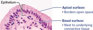
Organized Assembly: Arranged with distinct apical (top) and basolateral (bottom) surfaces.
Polarity: Apical surface faces open space; basal surface attaches to underlying connective tissue.
Highly Regenerative: Rapidly replaces damaged cells.
Avascular but Innervated: Lacks blood vessels but has nerve supply.
Classification of Epithelial Tissue
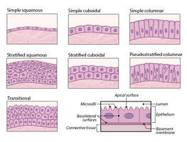
Epithelial tissues are classified by cell layers and cell shape:
Simple: Single layer of cells.
Stratified: Two or more layers.
Pseudostratified: Appears layered due to nuclei placement, but is a single layer.
Squamous: Flat, scale-like cells.
Cuboidal: Box-like cells.
Columnar: Tall, rectangular cells.
Types of Epithelia
Type | Description | Location |
|---|---|---|
Simple Squamous | Single layer of flat cells; filtration and diffusion | Air sacs of lungs, glomeruli, blood vessels, body cavity linings |
Simple Cuboidal | Single layer of cube-like cells; secretion and absorption | Ducts, glands, kidney tubules, ovary, thyroid |
Simple Columnar | Single layer of tall cells; absorption and secretion | Digestive and respiratory tract linings |
Pseudostratified Columnar | Single layer, nuclei at different heights; secretion and propulsion | Male sperm duct, respiratory tract, large glands |
Stratified Squamous | Multiple layers; protection against abrasion | Skin, esophagus, vagina, mouth |
Stratified Cuboidal/Columnar | Two or more layers; secretion | Sperm duct, sweat/mammary glands, pharynx, urethra |
Transitional | Stratified appearance; stretch permitted | Urinary tract: ureter, bladder, urethra |

Simple Squamous Epithelium
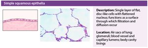
Description: Single layer of flat, disc-like cells with flattened nuclei; surface for filtration and diffusion.
Location: Air sacs of lung, glomeruli, blood vessel and capillary lumens, body cavity linings.
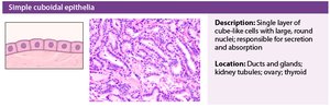
Simple Cuboidal Epithelium
Description: Single layer of cube-like cells with large, round nuclei; responsible for secretion and absorption.
Location: Ducts and glands, kidney tubules, ovary, thyroid.
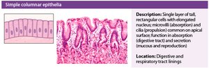
Simple Columnar Epithelium
Description: Single layer of tall, rectangular cells with elongated nuclei; microvilli and cilia common on apical surface; absorption and secretion.
Location: Digestive tract lining, respiratory tract lining.
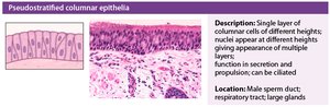
Pseudostratified Columnar Epithelium
Description: Single layer of columnar cells of different heights; nuclei at different heights; appearance of multiple layers; secretion and propulsion.
Location: Male sperm duct, respiratory tract, large glands.
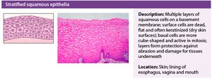
Stratified Squamous Epithelium
Description: Multiple layers of squamous cells; surface cells dead, flat, keratinized; basal cells cube-shaped and mitotically active; protection against abrasion.
Location: Skin, lining of esophagus, vagina, mouth.
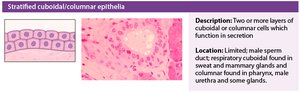
Stratified Cuboidal/Columnar Epithelium
Description: Two or more layers of cuboidal or columnar cells; function in secretion.
Location: Male sperm duct, sweat and mammary glands, pharynx, male urethra.
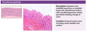
Transitional Epithelium
Description: Appears stratified squamous or cuboidal; basal cells cuboidal, surface cells rounded/squamous; stretch permitted for urine storage.
Location: Lining of urinary tract: ureter, bladder, urethra.
Glandular Epithelium
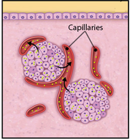
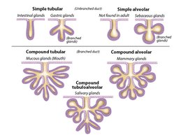
Glandular epithelium is classified as endocrine or exocrine based on where products are released.
Endocrine: Secrete hormones into localized capillaries.
Exocrine: Secrete products into ducts.
Functional Classification of Exocrine Glands
Type | Mechanism | Example |
|---|---|---|
Merocrine | Product secreted by exocytosis | Salivary glands |
Apocrine | Product secreted by pinching off membrane | Mammary glands |
Holocrine | Product secreted by cell rupture | Sebaceous glands |
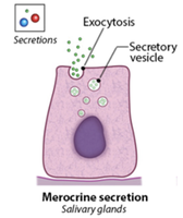
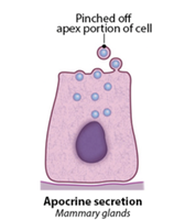
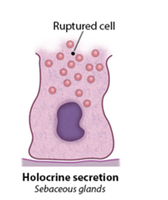
Connective Tissue
Overview and Functions
Connective tissue is the most abundant and widely distributed tissue type. It supports, protects, and binds other tissues and organs.
Protection: Shields tissues and organs.
Structural Support: Provides framework for muscle attachment and movement.
Energy Storage and Insulation: Adipose tissue stores energy and insulates.
Transport: Blood transports nutrients and gases.
Characteristics of Connective Tissue
Cells: Embedded in extracellular matrix.
Matrix: Composed of fibers and ground substance.
Vascularity: Varies from highly vascular (bone) to avascular (cartilage).
Origin: Derived from mesenchyme.

Cells and Fibers in Connective Tissue
Cells: Osteoblasts/cytes (bone), fibroblasts/cytes (proper), chondroblasts/cytes (cartilage), adipocytes (fat), blood cells (immunity).
Fibers: Collagen (strength), elastin (flexibility), reticulin (meshwork).
Muscle Tissue
Overview and Types
Muscle tissue produces movement, maintains posture, and generates heat. Muscle cells are called muscle fibers and require abundant ATP.
Skeletal Muscle: Voluntary movement; attached to bones; striated and multinucleated.
Cardiac Muscle: Involuntary; heart wall; striated with intercalated discs.
Smooth Muscle: Involuntary; walls of hollow organs; spindle-shaped, non-striated.
Nervous Tissue
Overview and Functions
Nervous tissue is specialized for communication and control. It detects stimuli and transmits electrical signals.
Neurons: Receive and transmit information; form synapses.
Glial Cells: Support, protect, and nourish neurons.
Body Membranes
Types of Membranes
Body membranes incorporate more than one tissue type and are classified as cutaneous, mucous, or serous.
Cutaneous: Skin; dry, exposed to air.
Mucous: Lines cavities open to exterior; wet, adapted for absorption and secretion.
Serous: Lines closed ventral cavities; allows sliding; pleurae, pericardium, peritoneum.
Tissue Repair
Steps of Tissue Repair
Inflammation: White blood cells and clotting factors enter site; clot forms.
Organization: Restoration of blood supply; fibroblasts lay down temporary matrix.
Regeneration and Fibrosis: Epithelium regenerates; scar tissue may form.
Developmental Aspects of Tissue
Embryonic Development
Three Primary Germ Layers: Ectoderm, mesoderm, endoderm.
These layers specialize to form all primary tissues and organs.
References
Marieb EN, Hoehn K. Chapter 4: Tissue: The Living Fabric. In: Marieb EN, Hoehn K, eds. Human Anatomy & Physiology. 12th ed. Pearson; 2025.
Redden J, Crivello J. Chapter 3: Histology. In: Anatomy & Physiology in Context. 1st ed. Top Hat; 2024.
