 뒤로
뒤로Histology: Epithelial and Connective Tissue Classification
스터디 가이드 - 스마트 노트
 자료에 맞춘 맞춤형 노트, 핵심 정의, 예시, 맥락을 확장해 제공합니다.
Histology: Tissue Types
Overview of Tissue Types
In human anatomy, tissues are collections of specialized cells and extracellular materials that perform specific functions. There are four major tissue types:
Epithelial tissue
Connective tissue
Muscle tissue (covered later in the semester)
Neural tissue (covered later in the semester)
Epithelial and connective tissues are foundational for understanding organ structure and function.
Epithelial Tissue Classification
General Characteristics
Epithelial tissue covers exposed surfaces, lines internal passageways and cavities, and produces glandular secretions. The basal lamina (basement membrane) anchors the epithelium to underlying connective tissue.
Cell Layers:
Simple: Single cell layer
Stratified: Two or more cell layers
Cell Shape:
Squamous: Thin, flat, irregularly shaped
Cuboidal: Cube-shaped, central nucleus
Columnar: Height greater than width, nuclei tend toward basal lamina
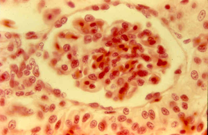
Simple Squamous Epithelium
Simple squamous epithelium consists of a single layer of flat cells. It allows for rapid absorption, secretion, filtration, and diffusion, and mitigates friction.
Locations:
Mesothelia of body cavities
Kidneys (blood filtration)
Alveoli of lungs (gas exchange)
Interior of blood vessels
Stratified Squamous Epithelium
Stratified squamous epithelium provides physical protection against mechanical abrasion and pathogens.
Locations:
Skin surface (keratinized)
Lining of mouth, esophagus, rectum, anus, vagina (non-keratinized)
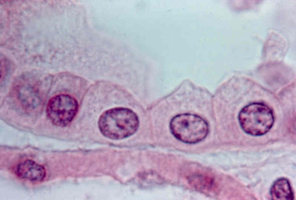
Simple Cuboidal Epithelium
Simple cuboidal epithelium is a single layer of cube-shaped cells. It is specialized for secretion and absorption, with limited protection.
Locations:
Ducts and glands
Parts of renal tubules
Stratified Cuboidal Epithelium
Stratified cuboidal epithelium provides protection, secretion, and absorption. It is not very common.
Locations:
Some ducts (e.g., sweat glands)
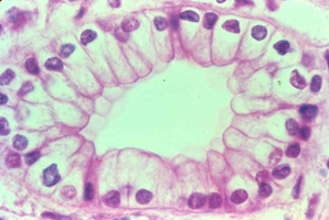
Simple Columnar Epithelium
Simple columnar epithelium consists of tall, column-shaped cells. It is specialized for secretion and absorption, often with microvilli to maximize surface area (brush border). It also provides protection.
Locations:
Lining of stomach, intestine, gallbladder, uterine tubes, collecting ducts of kidneys
Excretory ducts
Stratified Columnar Epithelium
Stratified columnar epithelium provides protection and is not very common.
Locations:
Salivary gland ducts
Mammary glands
Parts of the pharynx, epiglottis, anus, and urethra
Pseudostratified Columnar Epithelium
Pseudostratified columnar epithelium appears stratified but all cells contact the basal lamina. It provides protection and secretion. Ciliated forms act as a "mucus elevator."
Locations:
Nasal cavity, trachea, bronchi
Parts of male reproductive tract
Transitional Epithelium
Transitional epithelium allows organs to stretch and become distended. It is stratified; basal layers appear cuboidal/columnar, superficial layers appear cuboidal when relaxed and squamous when stretched.
Locations:
Urinary bladder
Connective Tissue Classification
General Characteristics
Connective tissue fills internal spaces, provides structural support, and stores energy. The extracellular matrix consists of fibers and ground substance (fluid, gel, or solid).
Collagen fibers: Strong, insoluble, made of collagen protein
Elastic fibers: Stretch and rebound, made of elastin protein
Reticular fibers: Fine mesh, made of collagen protein
Loose Areolar Connective Tissue
Loose areolar connective tissue cushions organs and provides flexible support. It includes immune cells such as macrophages and mast cells.
Locations:
Basal lamina
Around/supporting blood vessels, nerves, joints, organs
Components:
Fibroblasts/fibrocytes (cells)
Elastic fibers (thin, stain darker)
Collagen fibers (thick, stain lighter)
Gelatinous ground substance
Adipose Connective Tissue
Adipose connective tissue provides shock absorption, padding, insulation, and energy storage.
Locations:
Subcutaneous fat
Around eyes, kidneys
Components:
Adipocytes (cells)
Large lipid droplets
Nucleus and other organelles in margins
Reticular Connective Tissue
Reticular connective tissue provides a supportive internal framework and acts as a filter in lymphoid organs.
Locations:
Lymphoid organs (liver, spleen, lymph nodes, thymus)
Bone marrow
Components:
Fibroblasts/fibrocytes (cells)
Lymphocytes (white blood cells)
Reticular fibers (very dark staining)
Dense Regular Connective Tissue
Dense regular connective tissue binds bones and/or muscles together and reduces friction. It has a regular pattern of parallel collagen fibers.
Locations:
Tendons (muscle to bone)
Ligaments (bone to bone)
Deep fascia
Components:
Fibroblast/fibrocyte nuclei
Dense Irregular Connective Tissue
Dense irregular connective tissue resists forces applied from many directions and prevents overexpansion of organs.
Locations:
Capsules of visceral organs
Periosteum and perichondrium
Reticular layer of dermis
Components:
Fibroblasts/fibrocytes (cells)
Network of collagen fibers
Elastic Connective Tissue
Elastic connective tissue allows expansion and contraction of organs and blood vessels. True elastic connective tissue is not very common.
Locations:
Blood vessels
Bronchial tubes
Components:
Layers of wavy elastic fibers
Fibroblast/fibrocyte nuclei
Additional info:
Muscle and neural tissues will be covered in later chapters.
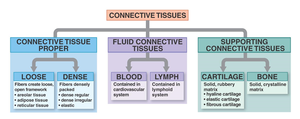
Connective tissue classification includes proper, fluid, and supporting types, as shown in the included diagram.
