 뒤로
뒤로Histology Part 2 & Integumentary System: Study Guide for Bio 223 Lab 2
스터디 가이드 - 스마트 노트
 자료에 맞춘 맞춤형 노트, 핵심 정의, 예시, 맥락을 확장해 제공합니다.
Histology Part 2: Connective and Nervous Tissues
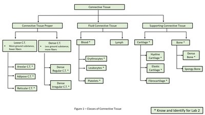
Overview of Connective Tissue
Connective tissue is the most diverse and abundant tissue class in the human body. It is characterized by a small number of cells surrounded by a substantial extracellular matrix, which consists of protein fibers and ground substance. The matrix may be solid, semi-solid, gelatinous, or fluid, depending on the tissue type.
Protein fibers: Reticular (fine, branching), Collagen (strong, rope-like), Elastic (stretchable and recoiling)
Cell types: Fibroblasts (produce matrix), mast cells, macrophages, mesenchymal cells, white blood cells
Categories: Connective tissue proper, supporting connective tissue, fluid connective tissue
Example: Connective tissue proper connects tissues and forms internal structures of organs.
Connective Tissue Proper
Loose Connective Tissue
Loose connective tissue contains fewer fibers that are widely spaced. It provides support and flexibility.
Areolar Connective Tissue: Irregular arrangement of fibers, withstands forces from multiple directions. Body locations: Between muscles, surrounding blood vessels and nerves, within the dermis.
Adipose Connective Tissue: Contains adipocytes specialized for lipid storage. Most cells appear empty due to slide preparation. Body locations: Hypodermis, surrounding eyeballs and kidneys, yellow bone marrow.
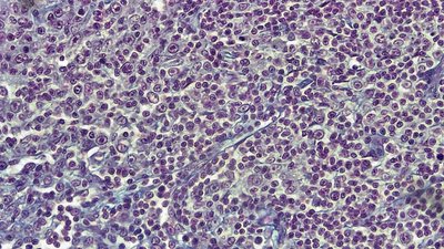
Reticular Connective Tissue: 3-D network of reticular fibers forms a stroma for support. Body locations: Kidney, spleen, lymph nodes, red bone marrow, liver.
Dense Connective Tissue
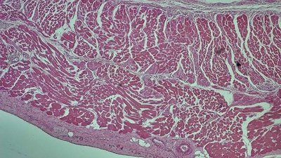
Dense Regular Connective Tissue: Densely packed collagen fibers oriented in the same direction; fibroblasts scattered between fibers. Body locations: Tendons, aponeuroses, most ligaments.
Dense Irregular Connective Tissue: Densely packed collagen fibers arranged irregularly, withstands pulling forces from many directions. Body locations: Dermis, periosteum, perichondrium, organ capsules.
Supporting Connective Tissue
Cartilage
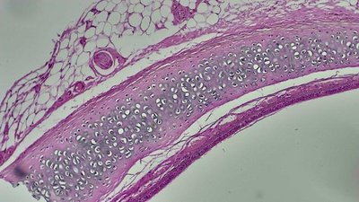
Hyaline Cartilage: Dense network of collagen fibers in gel-like matrix; chondrocytes in lacunae. Collagen fibers not visible. Body locations: Larynx, trachea, fetal skeleton, articular cartilage.
Elastic Cartilage: Numerous elastic fibers, resilient and flexible; chondrocytes in lacunae. Body locations: Auricle of the ear, epiglottis.
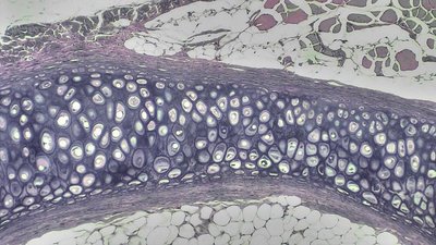
Fibrocartilage: Numerous bundles of collagen fibers visible in matrix; chondrocytes in lacunae. Provides tensile strength. Body locations: Pubic symphysis, intervertebral discs, meniscus of knee.
Bone
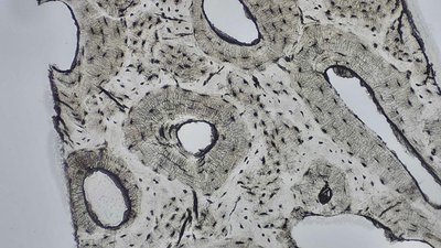
Dense Bone: Contains osteons (Haversian systems) with central canals for blood vessels and nerves; osteocytes in lacunae. Body locations: Bones of the skeleton.
Fluid Connective Tissue
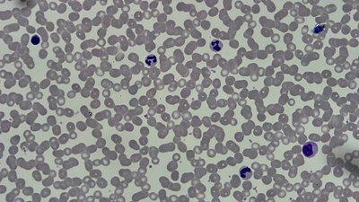
Blood: Fluid matrix (plasma) with formed elements: erythrocytes (red blood cells), leukocytes (white blood cells), platelets. Body locations: Chambers of the heart, blood vessels.
Nervous Tissue
Structure and Function
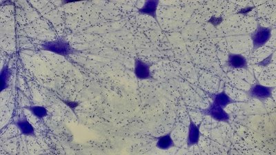
Nervous tissue consists of neurons and supporting cells. Neurons have three main components: perikaryon (cell body), axons, and dendrites. The perikaryon contains the nucleus and typical organelles. Axons and dendrites extend from the cell body and transmit electrical signals.
Body locations: Nervous system (brain, spinal cord, nerves)
Microscope slides: Multipolar neuron
Integumentary System
Anatomy of the Integumentary System
The integumentary system includes the skin and accessory structures. The skin is composed of the epidermis (keratinized, stratified squamous epithelium) and the dermis (connective tissue). The hypodermis is a subcutaneous layer deep to the dermis, consisting of adipose and areolar connective tissues. Accessory structures include hair, nails, sweat glands, sebaceous glands, and sensory receptors.
Epidermis layers: Stratum corneum, stratum lucidum, stratum granulosum, stratum spinosum, stratum basale
Dermis: Dermal papillae
Hypodermis: Adipose and areolar connective tissue
Tactile receptors: Lamellated (Pacinian) corpuscles, tactile (Meissner’s) corpuscles
Sweat glands: Apocrine and eccrine sweat glands
Hair follicle: Hair shaft (cortex, medulla, cuticle), hair root, hair bulb, hair papilla
Arrector pili muscle
Sebaceous glands
Integumentary System – Histology
Microscope slides of skin sections are used to identify the histological structures of the integumentary system. Key structures include sweat glands, hair follicle components, arrector pili muscle, sebaceous glands, and the layers of the epidermis and dermis.
Sweat glands: Apocrine and eccrine
Hair follicle: Hair shaft, hair bulb
Arrector pili muscle
Sebaceous glands
Epidermis layers: Stratum corneum, stratum lucidum, stratum granulosum, stratum spinosum, stratum basale
Dermis: Dermal papillae
Hypodermis
Summary Table: Classes of Connective Tissue
The following table summarizes the main classes and subtypes of connective tissue, as presented in Lab 2:
Class | Subtype | Example Tissue |
|---|---|---|
Connective Tissue Proper | Loose | Areolar, Adipose, Reticular |
Connective Tissue Proper | Dense | Dense Regular, Dense Irregular |
Supporting Connective Tissue | Cartilage | Hyaline, Elastic, Fibrocartilage |
Supporting Connective Tissue | Bone | Dense Bone, Spongy Bone |
Fluid Connective Tissue | Blood | Erythrocytes, Leukocytes, Platelets |
Fluid Connective Tissue | Lymph | --- |
Key Points for Lab Identification and Quiz Preparation
Define all bold terms from the lab manual.
Classify each tissue by its general type (connective or nervous).
Identify and label the 10 connective tissues and nervous tissue, using complete tissue names.
Provide one detailed body location for each tissue, as listed in the lab manual.
Identify integumentary system anatomy and histological structures on models and slides.
Use lab manual definitions and terms exclusively.
Additional info: For exam preparation, focus on spelling, complete tissue names, and detailed locations as specified in the lab manual.
