뒤로
뒤로Integumentary System, Bone Histology, and Muscle Structure: Study Notes
스터디 가이드 - 스마트 노트
 자료에 맞춘 맞춤형 노트, 핵심 정의, 예시, 맥락을 확장해 제공합니다.
Integumentary System
Skin Regions
The skin is composed of multiple layers, each with distinct structures and functions. It serves as the body's primary barrier against the environment and plays a role in sensation, temperature regulation, and protection.
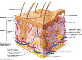
Epidermis: The outermost layer, composed mainly of keratinized stratified squamous epithelium. It provides a waterproof barrier and creates our skin tone.
Dermis: Located beneath the epidermis, this layer contains connective tissue, blood vessels, nerve endings, hair follicles, and glands. It is divided into the papillary and reticular layers.
Papillary Layer: The superficial layer of the dermis, consisting of areolar connective tissue with fingerlike projections called dermal papillae.
Reticular Layer: The deeper, thicker layer of the dermis, composed of dense irregular connective tissue.
Hypodermis (Subcutaneous Layer): Not technically part of the skin, this layer consists mainly of adipose tissue and anchors the skin to underlying structures.
Skin Structures
Several specialized structures are embedded within the skin, each contributing to its function.
Hair Shaft: The visible part of the hair above the skin surface.
Tactile (Meissner’s) Corpuscle: Sensory receptors for light touch, located in the dermal papillae.
Merkel Disc: Sensory receptors for light touch, found at the epidermal-dermal junction.
Sebaceous Gland: Produces sebum (oil) to lubricate skin and hair.
Arrector Pili Muscle: Smooth muscle attached to hair follicles; contraction causes "goosebumps."
Eccrine (Merocrine) Sweat Glands: Widely distributed glands that secrete sweat for thermoregulation.
Hair Follicle: The structure in the dermis from which hair grows.
Lamellar (Pacinian) Corpuscle: Deep pressure and vibration receptors in the dermis and hypodermis.
Nerve Fibers: Transmit sensory information from the skin to the central nervous system.
Epidermal Layers
The epidermis is organized into distinct layers, each with specialized cells and functions. In thick skin (palms, soles), an additional layer is present.
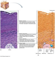
Stratum Corneum: Outermost layer; consists of dead, keratinized cells that provide a tough, protective barrier.
Stratum Lucidum: Present only in thick skin; a thin, translucent layer of dead cells.
Stratum Granulosum: Cells here contain granules that help form a waterproof barrier.
Stratum Spinosum: Several layers of keratinocytes connected by desmosomes; provides strength and flexibility.
Stratum Basale (Stratum Germinativum): Deepest layer; a single row of stem cells that continually divide to produce new keratinocytes.
Nail Structures
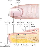
Nails are protective coverings on the dorsal surface of the distal fingers and toes. They are composed of hard keratin.
Free Edge: The part of the nail that extends beyond the finger or toe.
Eponychium (Cuticle): The fold of skin at the base of the nail plate.
Nail Body (Plate): The visible, attached part of the nail.
Nail Bed: The skin beneath the nail plate.
Lunule: The whitish, crescent-shaped area at the base of the nail.
Hyponychium: The area under the free edge of the nail.
Nail Root: The proximal part of the nail embedded under the skin.
Lateral and Proximal Folds: Skin folds that protect the sides and base of the nail.
Matrix: The region where nail growth occurs.
Hair Structures
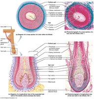
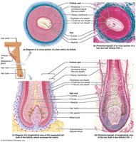
Hair is composed of dead, keratinized cells and serves protective and sensory functions. Each hair has several anatomical regions and associated structures.
Hair Shaft: The portion of hair above the skin surface.
Hair Root: The portion of hair below the skin surface.
Hair Bulb: The expanded base of the hair follicle.
Hair Matrix: The actively dividing area of the hair bulb that produces hair.
Hair Papilla: A dermal tissue that supplies nutrients to the growing hair.
Hair Follicle Wall: Composed of several layers, including the peripheral connective tissue sheath, glassy membrane, and epithelial root sheaths.
Cuticle, Cortex, Medulla: Layers of the hair shaft; the cuticle is the outermost, the cortex is the middle, and the medulla is the innermost layer.
Cutaneous Glands
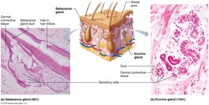
The skin contains several types of glands that contribute to its protective and regulatory functions.
Sebaceous (Oil) Glands: Secrete sebum into hair follicles to lubricate and waterproof the skin and hair.
Sweat (Sudoriferous) Glands: Two main types: eccrine (merocrine) glands, which are involved in thermoregulation, and apocrine glands, which are found in specific areas and become active at puberty.
Bone Histology and Structure
Bone Histology
Bones are dynamic organs composed of several tissue types. Their microscopic structure is essential for understanding their function and strength.
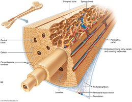
Periosteum: A dense layer of vascular connective tissue enveloping the bones except at the surfaces of the joints.
Endosteum: A thin vascular membrane lining the inner surface of the bony tissue.
Circumferential Lamellae: Layers of bony matrix that extend around the entire circumference of the shaft.
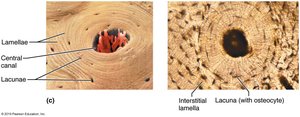
Interstitial Lamellae: Incomplete lamellae found between osteons.
Concentric Lamellae: Layers of bony matrix around a central canal in an osteon.
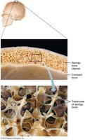
Trabeculae of Spongy Bone
Spongy bone is composed of a network of trabeculae, which provide structural support and house bone marrow.
Trabeculae: Thin columns and plates of bone that create a spongy structure, reducing bone weight while maintaining strength.
Osteon (Haversian) System
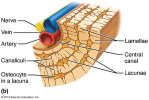
The osteon is the fundamental functional unit of compact bone, providing strength and facilitating nutrient delivery.
Osteon: Cylindrical structures that contain a central canal surrounded by concentric lamellae.
Lacunae: Small spaces between lamellae that house osteocytes.
Central (Haversian) Canal: Contains blood vessels and nerves.
Perforating (Volkmann’s) Canal: Channels that connect central canals of adjacent osteons.
Bone Structure
Long bones have a characteristic structure that supports their function in movement and support.
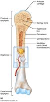
Proximal and Distal Epiphysis: The ends of the bone, usually wider than the shaft, and covered with articular cartilage.
Epiphyseal Plate/Line: The growth plate (cartilage) in children, which becomes the epiphyseal line (bone) in adults.
Metaphysis: The region between the diaphysis and epiphysis.
Diaphysis: The shaft of the bone, composed mainly of compact bone.
Medullary Cavity: The central cavity of bone shafts where marrow is stored.
Muscle Layers and Fiber Structure
Muscle Layers
Skeletal muscle is organized into layers of connective tissue that support and protect muscle fibers.
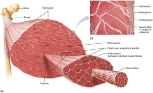
Epimysium: The outermost layer, surrounding the entire muscle.
Perimysium: Surrounds bundles of muscle fibers called fascicles.
Endomysium: Surrounds individual muscle fibers within a fascicle.
Muscle Fiber Subcellular Structures
Muscle fibers contain specialized structures that enable contraction and efficient energy use.
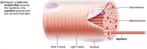
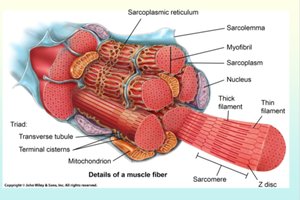
Sarcolemma: The plasma membrane of a muscle fiber.
Myofibril: Long, rod-like organelles within muscle fibers, composed of repeating sarcomeres.
Sarcoplasm: The cytoplasm of a muscle fiber, containing organelles and myofibrils.
Terminal Cistern: Enlarged areas of the sarcoplasmic reticulum that store calcium ions.
Sarcoplasmic Reticulum: Specialized endoplasmic reticulum that regulates calcium ion concentration.
Transverse Tubules (T-tubules): Invaginations of the sarcolemma that help transmit action potentials into the muscle fiber.
Thick Filament: Composed mainly of myosin; involved in muscle contraction.
Thin Filament: Composed mainly of actin; interacts with myosin for contraction.