 뒤로
뒤로Introduction to Anatomy: Anatomical Terminology, Body Cavities, and Organ Systems
스터디 가이드 - 스마트 노트
 자료에 맞춘 맞춤형 노트, 핵심 정의, 예시, 맥락을 확장해 제공합니다.
The Language of Anatomy
Anatomic Position
The anatomic position is a standardized posture used as a reference in describing the relationship of body parts. It is essential for clear communication in anatomy.
Upright stance
Feet parallel and flat on the floor
Upper limbs at the sides of the body
Palms face anteriorly (toward the front)
Head is level
Eyes look forward
Importance: The anatomic position provides a consistent frame of reference for anatomical terminology and descriptions.
Anatomical Regions and Surface Landmarks
Body regions are specific areas used to describe locations on the body. These regions help in identifying the position of structures and are essential for clinical and anatomical communication.
Cephalic (head), Cervical (neck), Thoracic (chest), Abdominal (abdomen), Pelvic (pelvis), Upper limb, Lower limb, Dorsum (back), etc.
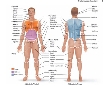
Additional info: Table 1.1 in most textbooks provides a comprehensive list of these regions.
Directional Terms
Directional terms are used to describe the locations of structures relative to other structures or locations in the body.
Anterior (ventral): Toward the front of the body
Posterior (dorsal): Toward the back of the body
Superior (cranial): Toward the top of the body
Inferior (caudal): Toward the bottom of the body
Medial: Toward the midline of the body
Lateral: Away from the midline of the body
Proximal: Closer to the point of attachment (used for limbs)
Distal: Farther from the point of attachment (used for limbs)
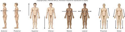
Note: Proximal and distal are only used when referring to limbs.
Sections and Planes
Sections and planes are used to describe how the body or an organ is cut to examine internal structures.
Section: An actual cut or slice that exposes internal anatomy
Plane: An imaginary flat surface passing through the body
Coronal (frontal) plane: Divides the body into anterior (front) and posterior (back) portions
Transverse (horizontal) plane: Divides the body into superior (top) and inferior (bottom) portions
Midsagittal plane: Divides the body into equal left and right halves
Sagittal plane: Divides the body into left and right portions (not necessarily equal)
Parasagittal plane: Divides the body into unequal left and right portions
Oblique plane: Divides the structure at an angle
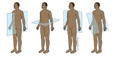
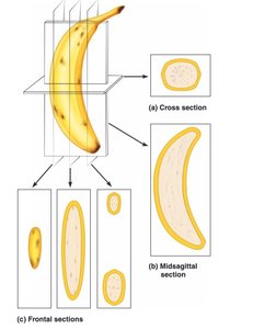
Example: A CT scan often uses the transverse plane to produce cross-sectional images of the body.
Body Cavities and Membranes
Major Body Cavities
The body contains several major cavities that house and protect internal organs.
Dorsal cavity: Includes the cranial cavity (brain) and vertebral canal (spinal cord)
Ventral cavity: Includes the thoracic cavity (heart and lungs) and abdominopelvic cavity (abdominal and pelvic organs)
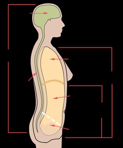
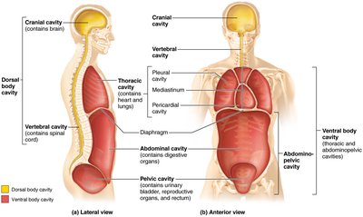
Example: The heart is located in the thoracic cavity, specifically within the mediastinum.
Serous Membranes
Serous membranes are thin, double-layered membranes that line the ventral body cavities and cover the organs within these cavities. They secrete serous fluid to reduce friction between moving organs.
Parietal layer: Lines the internal surface of the body wall
Visceral layer: Covers the external surface of organs
Serous cavity: Space between the layers, containing serous fluid
Examples of serous membranes:
Pericardium: Surrounds the heart
Pleura: Surrounds the lungs
Peritoneum: Surrounds most abdominal organs
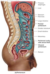
Note: The kidneys are retroperitoneal, meaning they are located behind the peritoneum.
Abdominopelvic Quadrants and Regions
Abdominopelvic Quadrants
The abdominopelvic cavity is divided into four quadrants for clinical reference:
Right Upper Quadrant (RUQ)
Left Upper Quadrant (LUQ)
Right Lower Quadrant (RLQ)
Left Lower Quadrant (LLQ)
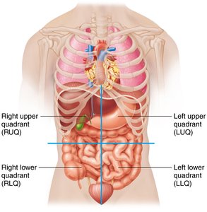
Example: The appendix is typically located in the RLQ.
Abdominopelvic Regions
For more precise anatomical study, the abdominopelvic cavity is divided into nine regions:
Right hypochondriac
Epigastric
Left hypochondriac
Right lumbar
Umbilical
Left lumbar
Right inguinal (iliac)
Hypogastric (pubic)
Left inguinal (iliac)
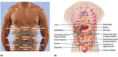
Example: The liver is primarily in the right hypochondriac and epigastric regions.
Overview of Human Organ Systems
Introduction to Organ Systems
The human body is organized into eleven major organ systems, each with specific functions and major organs. Understanding these systems is foundational for the study of anatomy and physiology.
Integumentary System: Provides protection, prevents water loss/gain, synthesizes vitamin D, regulates temperature, and houses sensory receptors.
Skeletal System: Provides support and protection, site of blood cell production, stores minerals, and provides attachment for muscles and ligaments.
Muscular System: Produces movement and generates heat.
Nervous System: Regulatory system, responds to stimuli, controls muscles and glands, responsible for consciousness, intelligence, and memory.
Endocrine System: Produces hormones, regulates homeostasis, controls digestive and reproductive processes.
Cardiovascular System: Pumps and transports blood, distributes nutrients, gases, hormones, and removes waste products.
Lymphatic System: Transports and filters lymph, participates in immune responses.
Respiratory System: Responsible for gas exchange between air, blood, and lungs.
Urinary System: Filters blood, removes waste, concentrates urine, and expels it from the body.
Digestive System: Digests food, absorbs nutrients, and expels waste.
Reproductive System: Produces sex cells and hormones.
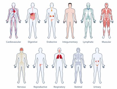
Example: The cardiovascular system includes the heart and blood vessels, which work together to circulate blood throughout the body.
Summary Table: Human Organ Systems
System | Major Organs | Primary Functions |
|---|---|---|
Integumentary | Skin, hair, nails | Protection, temperature regulation, sensory reception |
Skeletal | Bones, joints | Support, protection, blood cell production, mineral storage |
Muscular | Muscles | Movement, heat production |
Nervous | Brain, spinal cord, nerves | Regulation, response to stimuli, cognition |
Endocrine | Glands (pituitary, thyroid, etc.) | Hormone production, homeostasis |
Cardiovascular | Heart, blood vessels | Transport of blood, nutrients, gases, wastes |
Lymphatic | Lymph nodes, lymphatic vessels | Immune response, fluid balance |
Respiratory | Lungs, trachea | Gas exchange |
Urinary | Kidneys, bladder | Waste removal, water balance |
Digestive | Stomach, intestines, liver | Digestion, nutrient absorption, waste elimination |
Reproductive | Ovaries/testes, uterus/prostate | Production of sex cells, reproduction |
Additional info: For more details, refer to Table 2.1 in your textbook.
