 뒤로
뒤로Introduction to Anatomy & Physiology: Core Concepts and Organization
스터디 가이드 - 스마트 노트
 자료에 맞춘 맞춤형 노트, 핵심 정의, 예시, 맥락을 확장해 제공합니다.
Introduction to Anatomy and Physiology
Definition and Scope
Anatomy and Physiology (A&P) are foundational sciences for understanding the structure and function of the human body. Anatomy is the study of the structure and physical relationships of body parts, while Physiology focuses on the functions and processes of those parts. Together, they provide a comprehensive understanding of how the body operates and maintains life.
Characteristics of Living Things
Cellular Composition: All living organisms are composed of one or more cells.
Metabolism: The sum of all chemical reactions that occur within the body.
Growth: Increase in size and number of cells.
Excretion: Removal of waste products from the body.
Responsiveness: Ability to sense and react to changes in the environment.
Movement: Motion of the whole body, organs, cells, or organelles.
Reproduction: Production of new cells or organisms.

Levels of Structural Organization
Hierarchy of Organization
The human body is organized into a series of hierarchical levels, each building upon the previous, similar to a set of Russian dolls. This organization allows for increasing complexity and specialization.
Chemical Level: Atoms and molecules essential for life.
Cellular Level: Cells are the basic units of life.
Tissue Level: Groups of similar cells performing a common function.
Organ Level: Structures composed of two or more tissue types working together.
Organ System Level: Groups of organs that perform related functions.
Organism Level: The complete living being.

Specialized Subfields in Anatomy & Physiology
Classification of Subfields
Subfields in anatomy are typically classified by the size and visibility of the structures studied:
Gross Anatomy: Study of structures visible to the naked eye.
Microscopic Anatomy: Study of structures requiring a microscope, including:
Histology: Study of tissues.
Cytology: Study of cells.
Physiology subfields are classified by organ system (e.g., neurophysiology, cardiovascular physiology).
Language of Anatomy & Anatomical Position
Standard Terminology and Position
To ensure clear communication, anatomy uses standardized terms, often derived from Latin or Greek. The anatomical position is the common reference point: standing upright, facing forward, arms at sides, palms facing forward.
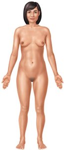
Directional Terms
Term | Definition | Example |
|---|---|---|
Anterior (ventral) | Toward the front | The palms are on the anterior side of the body. |
Posterior (dorsal) | Toward the back | The spinal cord is posterior to the esophagus. |
Superior (cranial) | Toward the head | The nose is superior to the mouth. |
Inferior (caudal) | Toward the tail | The umbilicus is inferior to the chest. |
Proximal | Closer to the point of origin | The knee is proximal to the ankle. |
Distal | Farther from the point of origin | The wrist is distal to the elbow. |
Medial | Closer to the midline | The ear is medial to the shoulder. |
Lateral | Farther from the midline | The shoulder is lateral to the chest. |
Superficial | Closer to the surface | The skin is superficial to the muscle. |
Deep | Farther below the surface | Bone is deep to the skin. |
Regional Anatomy
Axial Region: Head, neck, and trunk.
Appendicular Region: Limbs and girdles.
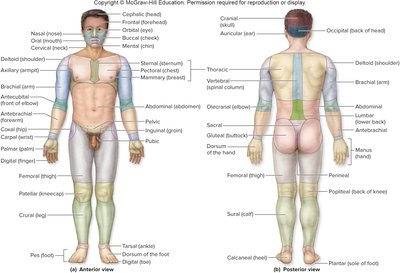
Planes of Section
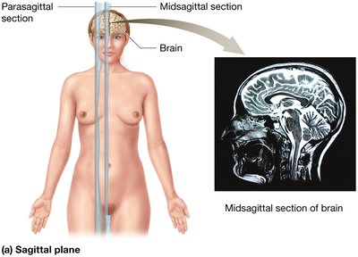
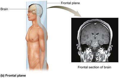
Body sections are described using standard anatomical planes:
Sagittal Plane: Divides the body into right and left parts (midsagittal = equal halves).
Frontal (Coronal) Plane: Divides the body into anterior and posterior parts.
Transverse (Horizontal) Plane: Divides the body into superior and inferior parts.
Body Cavities and Membranes
Major Body Cavities
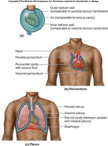
Body cavities are fluid-filled spaces that protect organs and allow for changes in organ shape and size. The axial region contains two main cavities:
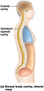
Dorsal (Posterior) Cavity: Contains the cranial and vertebral cavities.
Ventral (Anterior) Cavity: Contains the thoracic and abdominopelvic cavities.
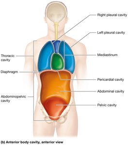
Posterior (Dorsal) Body Cavity
Cranial Cavity: Houses and protects the brain.
Vertebral (Spinal) Cavity: Encloses the spinal cord.
Both are filled with cerebrospinal fluid for protection.
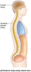
Anterior (Ventral) Body Cavity
Thoracic Cavity: Superior to the diaphragm; contains the lungs and heart.
Abdominopelvic Cavity: Inferior to the diaphragm; contains digestive, urinary, and reproductive organs.
The diaphragm separates the thoracic and abdominopelvic cavities.
Thoracic Cavity Subdivisions
Pleural Cavities: Surround each lung.
Mediastinum: Central compartment containing the heart, great vessels, trachea, and esophagus.
Pericardial Cavity: Surrounds the heart.
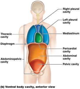
Abdominopelvic Cavity Subdivisions
Abdominal Cavity: Contains digestive organs.
Pelvic Cavity: Contains urinary and reproductive organs.
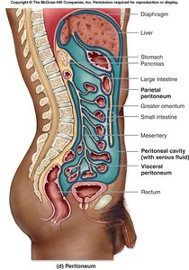
Peritoneal Cavity: Subcavity within the abdominal cavity, lined by the peritoneum.
Abdominopelvic Segmentation
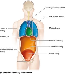
The abdominopelvic cavity can be divided for clinical and anatomical reference:
Quadrant System: Four quadrants (RUQ, LUQ, RLQ, LLQ) using two perpendicular lines at the umbilicus.
Nine-Region System: Nine regions using four lines (two parasagittal, two transverse).
Serous Membranes
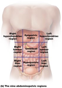
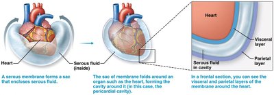
Serous membranes line body cavities and cover organs, reducing friction. They consist of two layers:
Visceral Layer: Closest to the organ.
Parietal Layer: Lines the cavity wall.
Between layers is serous fluid, which lubricates and protects organs.
Core Principles in Anatomy & Physiology
Homeostasis
Homeostasis is the maintenance of a stable internal environment. Disruption of homeostasis can lead to disease or death if not corrected.
Feedback Loops
Negative Feedback Loops: Oppose initial changes and reduce output to maintain set points (e.g., body temperature regulation).
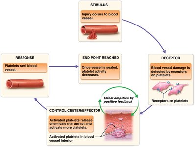
Positive Feedback Loops: Reinforce initial changes and amplify output, often to expedite a process (e.g., blood clotting, labor contractions, lactation).
Principle of Form and Function
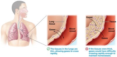
Structure and function are closely related in biology. The shape of a structure often determines its function, and vice versa. For example, the thin walls of lung tissue facilitate rapid gas exchange.
Principle of Gradients
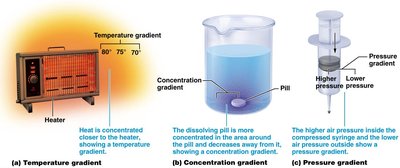
Gradients—differences in concentration, pressure, or temperature—drive many physiological processes, such as diffusion and osmosis.
Principle of Cell Communication
Cells communicate via electrical and chemical signals to coordinate functions and maintain homeostasis. For example, nerve cells release neurotransmitters to stimulate muscle contraction.
Additional info: Where content was incomplete, standard academic definitions and examples were provided to ensure clarity and completeness for college-level Anatomy & Physiology students.
