 뒤로
뒤로Introduction to Anatomy and Physiology: Core Concepts and Structural Organization
스터디 가이드 - 스마트 노트
 자료에 맞춘 맞춤형 노트, 핵심 정의, 예시, 맥락을 확장해 제공합니다.
Introduction to Anatomy and Physiology
Overview of Anatomy and Physiology
Anatomy and physiology are foundational sciences for understanding the structure and function of the human body. Anatomy focuses on the physical structures, while physiology examines the processes and functions that sustain life.
Anatomy: The study of body structure, including organs, tissues, and cells.
Physiology: The study of how the body and its parts function.
Applications: Essential for healthcare, diagnostics, and understanding disease mechanisms.
Characteristics of Living Organisms
Key Properties Shared by Living Organisms
All living organisms exhibit several distinct properties that define life and distinguish them from non-living matter.
Cellular Composition: Cells are the basic units of life; all organisms are composed of cells.
Metabolism: The sum of all chemical reactions in the body, including anabolic (building up) and catabolic (breaking down) processes.
Growth: Increase in size of individual cells or increase in number of cells.
Excretion: Removal of waste products generated by metabolic processes.
Responsiveness (Irritability): Ability to sense and respond to environmental changes or stimuli.
Movement: Movement of the organism as a whole, individual cells, or materials within cells.
Reproduction: Production of new cells (cellular reproduction) and new organisms (organismal reproduction).
Levels of Structural Organization and Body Systems
Hierarchical Organization of the Human Body
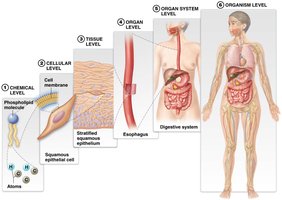
The human body is organized into six structural levels, each building upon the previous, from the simplest chemical level to the complex organism level.
Chemical Level: Atoms and molecules form the basis of all matter.
Cellular Level: Cells are formed from molecules and are the basic units of life.
Tissue Level: Groups of similar cells performing a common function (e.g., epithelial tissue).
Organ Level: Structures composed of two or more tissue types (e.g., esophagus).
Organ System Level: Groups of organs working together to perform major functions (e.g., digestive system).
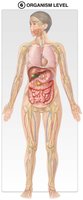
Organism Level: All organ systems functioning together to form the complete human body.
Types of Anatomy and Physiology
Approaches to Studying the Human Body
Anatomy and physiology can be studied using various approaches, each providing unique insights into structure and function.
Systemic Anatomy: Study of individual organ systems.
Regional Anatomy: Study of specific regions of the body (e.g., head, neck).
Surface Anatomy: Study of surface markings and features.
Gross Anatomy: Study of structures visible to the naked eye.
Microscopic Anatomy: Study of structures requiring a microscope, including histology (tissues) and cytology (cells).
Physiology Subfields: Includes neurophysiology (brain and nerves), cardiovascular physiology (heart and blood vessels), and others.
The Language of Anatomy and Physiology
Scientific Terminology and Word Roots
Anatomical and physiological terms are constructed from word roots, prefixes, and suffixes, enabling precise communication in science and medicine.
Word Roots: Core components of scientific terms.
Prefixes and Suffixes: Modify word roots to specify meaning.
Example: "Cardio-" (heart), "-itis" (inflammation), "carditis" (inflammation of the heart).
Anatomical Position
Standard Reference for Describing the Human Body
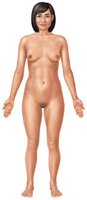
The anatomical position is a universally accepted reference posture for describing locations and relationships of body parts.
Definition: Standing upright, feet shoulder-width apart, arms at sides, palms facing forward.
Importance: Ensures consistency in anatomical descriptions.
Right and Left: Always refer to the subject's right and left, not the observer's.
Directional Terms
Describing Relative Locations in the Body
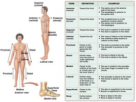
Directional terms are used to describe the positions of structures relative to each other, ensuring clear and accurate communication.
Common Terms: Anterior (ventral), posterior (dorsal), superior, inferior, proximal, distal, medial, lateral, superficial, deep.
Application: Used in clinical and anatomical descriptions.
Term | Definition | Example |
|---|---|---|
Anterior (ventral) | Toward the front | The palms are on the anterior side of the body. |
Posterior (dorsal) | Toward the back | The spine is posterior to the chest. |
Superior | Toward the head | The nose is superior to the mouth. |
Inferior | Toward the feet | The stomach is inferior to the heart. |
Proximal | Closer to point of origin | The shoulder is proximal to the wrist. |
Distal | Farther from point of origin | The fingers are distal to the elbow. |
Medial | Closer to midline | The nose is medial to the eyes. |
Lateral | Farther from midline | The ears are lateral to the nose. |
Superficial | Closer to surface | The skin is superficial to the muscles. |
Deep | Farther from surface | Bone is deep to muscle. |
Planes of Section
Dividing the Body for Study and Examination
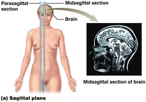
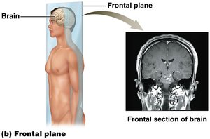
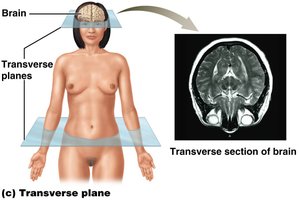
Planes of section are imaginary lines used to divide the body or its parts for anatomical study and medical imaging.
Sagittal Plane: Divides body into right and left sections; includes midsagittal (equal halves) and parasagittal (unequal halves).
Frontal (Coronal) Plane: Divides body into anterior and posterior sections.
Transverse (Horizontal) Plane: Divides body into superior and inferior sections.
Oblique Plane: Divides body at an angle; less commonly used.
Body Cavities
Major Cavities and Their Functions
Body cavities are fluid-filled spaces that house and protect internal organs, allowing them to move and expand as needed.
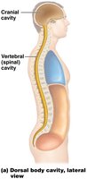
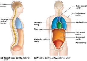
Dorsal Body Cavity: Located on the posterior side; includes cranial cavity (brain) and vertebral cavity (spinal cord).
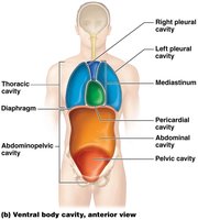
Ventral Body Cavity: Located on the anterior side; divided by the diaphragm into thoracic and abdominopelvic cavities.
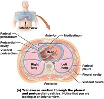
Thoracic Cavity: Contains pleural cavities (lungs), mediastinum (heart, trachea, esophagus), and pericardial cavity (heart).
Abdominopelvic Cavity: Contains abdominal (digestive organs) and pelvic (reproductive, urinary organs) cavities.
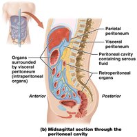
Peritoneal Cavity: Subdivision within the abdominal cavity, lined by serous membrane.
Abdominopelvic Quadrants and Regions
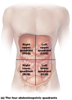
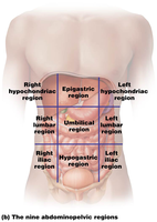
The abdominopelvic cavity can be divided into four quadrants or nine regions to aid in diagnosis and anatomical study.
Four Quadrants: Right upper (RUQ), right lower (RLQ), left upper (LUQ), left lower (LLQ).
Nine Regions: Right/left hypochondriac, epigastric, right/left lumbar, umbilical, right/left iliac, hypogastric.
Clinical Application: Helps localize pain and diagnose conditions based on organ location.
Serous Membranes
Structure and Function of Serous Membranes
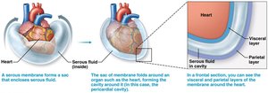
Serous membranes are thin sheets of tissue that line certain body cavities and surround organs, providing lubrication and reducing friction.
Layers: Visceral layer (contacts organ), parietal layer (attached to surrounding structures).
Serous Fluid: Lubricates and prevents friction between organ and cavity wall.
Main Serous Membranes: Pleural (lungs), pericardial (heart), peritoneal (abdominal organs).
Retroperitoneal Organs: Organs located behind the parietal peritoneum (e.g., kidneys).
Homeostasis and Feedback Mechanisms
Maintaining Internal Balance
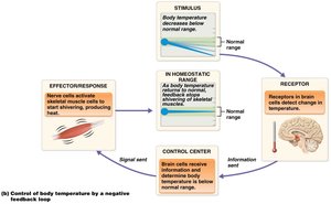
Homeostasis is the maintenance of a stable internal environment, essential for health and survival. It is regulated by feedback loops.
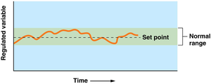
Regulated Variables: Variables maintained within a narrow range (e.g., temperature, blood glucose).
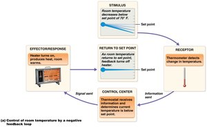
Negative Feedback: Opposes initial change, reduces output, and returns variable to set point.
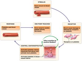
Positive Feedback: Amplifies initial change, reinforces stimulus, and shuts off when normal range is restored.
Misconceptions: Negative feedback is not "bad"; homeostasis is dynamic, not static; feedback loops are not simply "on" or "off"; only variables with receptors can be regulated.
Example: Childbirth and Positive Feedback
Childbirth is a classic example of a positive feedback loop, where the release of oxytocin causes uterine contractions, which further stimulate oxytocin release until delivery is complete.
Stimulus: Baby's head stretches cervix.
Control Center: Brain signals uterus.
Effector: Uterus produces oxytocin.
Response: Stronger contractions, more stretching, more oxytocin.
Pitocin: Synthetic oxytocin used to induce labor.
 Additional info: The notes have been expanded with academic context and examples to ensure completeness and clarity for exam preparation.
