 뒤로
뒤로Introduction to Anatomy and Physiology: Core Concepts and Structural Organization
스터디 가이드 - 스마트 노트
 자료에 맞춘 맞춤형 노트, 핵심 정의, 예시, 맥락을 확장해 제공합니다.
Introduction to Anatomy and Physiology
Overview of Anatomy and Physiology
Anatomy and physiology are foundational sciences in understanding the human body. Anatomy is the study of the structure of the human body, while physiology focuses on the functions of body parts. The core concept is that form and function are inextricably linked—each anatomical structure is designed to perform a specific physiological role.
Characteristics of Living Organisms
Cellular Composition: All living things are composed of cells, the basic units of life.
Metabolism: The sum of all chemical reactions in the body. Anabolism builds complex molecules, while catabolism breaks them down.
Growth: Increase in size and/or number of cells.
Excretion: Removal of metabolic waste products.
Responsiveness (Irritability): Ability to sense and react to environmental changes.
Movement: Motion of the organism or its parts.
Reproduction: Formation of new cells for growth, repair, or producing offspring.
Levels of Structural Organization
Six Structural Levels
The human body is organized into a hierarchy of structural levels, each building on the previous one:
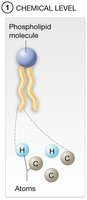
Chemical Level: Atoms and molecules form the foundation of all matter.
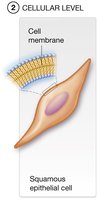
Cellular Level: Molecules combine to form cells, the smallest living units.
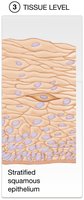
Tissue Level: Groups of similar cells and their extracellular matrix perform common functions.
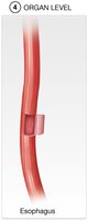
Organ Level: Two or more tissue types form organs with specific shapes and functions.
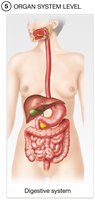
Organ System Level: Groups of organs work together to perform broad functions.
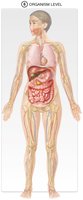
Organism Level: All organ systems function together to sustain life in the human body.
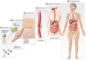
Examples of Structural Levels
Chemical Level: Phospholipid molecules and atoms (e.g., H, C).
Cellular Level: Squamous epithelial cell with a cell membrane.
Tissue Level: Stratified squamous epithelium.
Organ Level: Esophagus.
Organ System Level: Digestive system.
Organism Level: The complete human body.
The 11 Organ Systems of the Human Body
Overview of Organ Systems
The human body consists of 11 organ systems, each with specialized functions essential for survival and homeostasis.
Organ System | Main Functions |
|---|---|
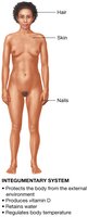
Integumentary | Protects body, regulates temperature, prevents water loss, produces vitamin D |
Skeletal | Supports and protects, stores calcium, produces blood cells |
Muscular | Produces movement, generates heat |
Nervous | Regulates body functions, sensation, movement, cognition |
Endocrine | Regulates body functions via hormones |
Cardiovascular | Transports blood, nutrients, wastes |
Lymphatic | Returns tissue fluid, immunity |
Respiratory | Gas exchange, acid-base balance |
Digestive | Digests food, absorbs nutrients, eliminates waste |
Urinary | Removes waste, fluid/electrolyte balance |
Reproductive | Produces gametes, sexual function |
Types of Anatomy and Physiology
Types of Anatomy
Systemic Anatomy: Study by organ systems.
Regional Anatomy: Study by body regions.
Surface Anatomy: Study of surface markings.
Gross Anatomy: Structures visible to the naked eye.
Microscopic Anatomy: Structures visible only with a microscope (includes histology and cytology).
Types of Physiology
Neurophysiology: Study of brain and nerves.
Cardiovascular Physiology: Study of heart and blood vessels.
Other subfields focus on chemical, cellular, tissue, or organ levels.
The Language of Anatomy and Physiology
Word Parts
Scientific terms are built from word roots, prefixes, and suffixes. For example, "anencephalic" means lacking a part of the brain (an- = without, encephala- = brain, -ic = condition of).
Anatomical Position
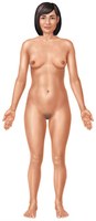
The anatomical position is the standard reference for describing body parts and regions:
Body standing upright
Feet shoulder-width apart
Arms at sides, palms facing forward
Head facing forward
"Right" and "left" refer to the subject's sides, not the observer's
Directional Terms
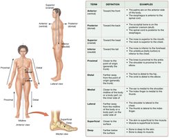
Directional terms describe the locations of body parts relative to each other. Common pairs include:
Term | Definition | Example |
|---|---|---|
Anterior (ventral) | Toward the front | The sternum is anterior to the heart |
Posterior (dorsal) | Toward the back | The heart is posterior to the sternum |
Superior (cranial) | Toward the head | The nose is superior to the mouth |
Inferior (caudal) | Toward the tail | The mouth is inferior to the nose |
Proximal | Closer to point of origin (limbs) | The elbow is proximal to the wrist |
Distal | Farther from point of origin (limbs) | The wrist is distal to the elbow |
Medial | Closer to midline | The heart is medial to the lungs |
Lateral | Farther from midline | The lungs are lateral to the heart |
Superficial | Closer to surface | The skin is superficial to muscles |
Deep | Farther from surface | The bones are deep to the skin |
Regional Terms
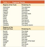
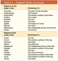
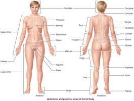
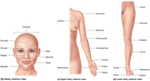
The body is divided into the axial region (head, neck, trunk) and the appendicular region (limbs). Each region is further subdivided and named for clarity in anatomical study.
Planes of Section
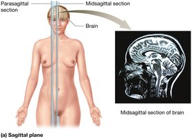
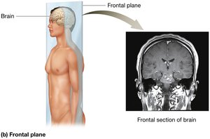
Three primary planes are used to divide the body for anatomical study:
Sagittal Plane: Divides body into right and left sections. Midsagittal is equal halves; parasagittal is unequal.
Frontal (Coronal) Plane: Divides body into anterior and posterior sections.
Transverse (Horizontal) Plane: Divides body into superior and inferior sections.
Oblique Plane: Divides body at an angle (less common).
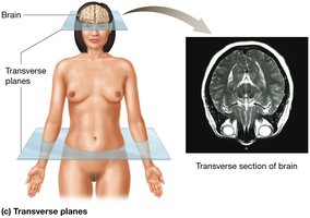
Organization of the Human Body
Body Cavities
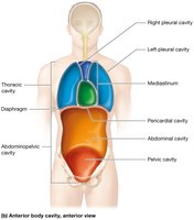
Body cavities are spaces within the body that protect organs and allow for movement and expansion. The axial region contains two main cavities:
Posterior Body Cavity: Cranial cavity (protects brain) and vertebral cavity (protects spinal cord), filled with cerebrospinal fluid.
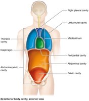
Anterior Body Cavity: Divided by the diaphragm into thoracic and abdominopelvic cavities, with smaller cavities formed by serous membranes.
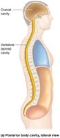
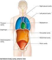
Thoracic and Abdominopelvic Cavities
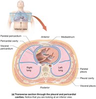
Thoracic Cavity: Contains pleural cavities (lungs), mediastinum (heart, trachea, esophagus), and pericardial cavity (heart).
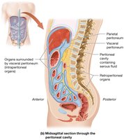
Abdominopelvic Cavity: Subdivided into abdominal (digestive organs) and pelvic (reproductive, urinary organs) cavities. The peritoneal cavity surrounds some abdominal organs.
Abdominopelvic Quadrants and Regions
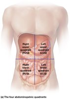
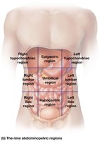
The abdominopelvic cavity can be divided for diagnostic purposes:
Four Quadrants: Right upper (RUQ), left upper (LUQ), right lower (RLQ), left lower (LLQ).
Nine Regions: Right/left hypochondriac, lumbar, iliac; epigastric, umbilical, hypogastric.
Serous Membranes
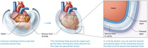
Serous membranes are thin sheets of tissue that form double layers around organs in the anterior body cavity. They secrete serous fluid to reduce friction from organ movement.
Visceral Layer: Contacts the organ.
Parietal Layer: Attaches to surrounding structures.
Serous Fluid: Lubricates and prevents friction.
Pleural Membranes: Surround the lungs.
Pericardial Membranes: Surround the heart.
Peritoneal Membranes: Surround some abdominal organs; retroperitoneal organs lie behind the parietal peritoneum.
Medical Imaging
Common Imaging Techniques
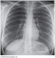
X-Ray: Uses ionizing radiation to visualize dense structures (e.g., bones, chest).
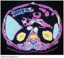
Computed Tomography (CT): Uses ionizing radiation and computer processing for 3D images, often in transverse sections.
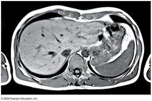
Magnetic Resonance Imaging (MRI): Uses magnetic fields and radio waves for detailed images, especially of soft tissues.
Core Principles in Anatomy and Physiology
Homeostasis
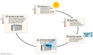
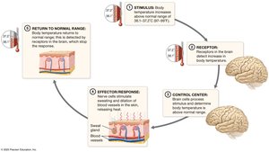
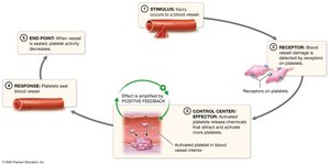
Homeostasis is the maintenance of a stable internal environment. Disturbances can lead to disease or death. Variables are regulated to stay near a set point, with feedback mechanisms maintaining balance.
Feedback Loops
Negative Feedback: Opposes changes, returning variables to normal (e.g., body temperature regulation).
Positive Feedback: Amplifies changes, often for a specific purpose (e.g., blood clotting, childbirth).
Structure and Function
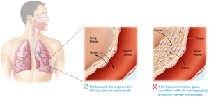
The principle of complementarity states that the form of a structure is always suited to its function. This applies at all levels, from molecules to organ systems.
Gradients
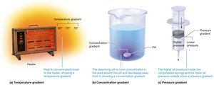
A gradient exists when more of something is present in one area than another, driving physiological processes such as respiration, nutrient exchange, and blood flow. Types include temperature, concentration, and pressure gradients.
Cell-Cell Communication
Cells communicate to coordinate body functions and maintain homeostasis. Communication occurs via electrical signals (e.g., nerve impulses) and chemical messengers (e.g., hormones).
Summary Table: Core Principles
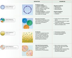
Core Principle | Definition | Examples |
|---|---|---|
Feedback Loops | Negative feedback opposes change; positive feedback amplifies change | Body temperature, blood clotting |
Structure-Function | Form suits function at all levels | Thin lung tissue for gas exchange |
Gradients | Difference in concentration, pressure, or temperature drives processes | Oxygen diffusion, blood flow |
Cell-Cell Communication | Electrical and chemical signals coordinate function | Nerve impulses, hormones |
