 뒤로
뒤로Introduction to Anatomy and Physiology: Foundations, Organization, and Homeostasis
스터디 가이드 - 스마트 노트
 자료에 맞춘 맞춤형 노트, 핵심 정의, 예시, 맥락을 확장해 제공합니다.
Introduction to Anatomy & Physiology
Definition and Scope
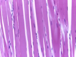
Anatomy is the study of the structures of the body, including what they are made of and where they are located. Physiology is the study of the functions of anatomical structures. The relationship between structure and function is fundamental; for example, the structure of skeletal muscle fibers enables their function in movement.
Anatomy: Focuses on body structures, their composition, and location.
Physiology: Explores how anatomical structures function individually and collectively.
Example: Skeletal muscle fibers are long, straight, and striated, allowing them to contract and move the skeleton.
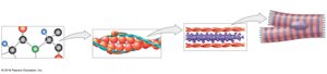
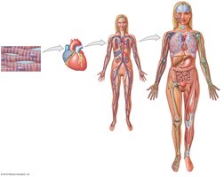
Levels of Organization in the Human Body
Chemical to Organism Level
The human body is organized into hierarchical levels, each building upon the previous:
Chemical Level: Atoms combine to form molecules, the building blocks of cells.
Cellular Level: Cells are the smallest living units, composed of molecules and organelles.
Tissue Level: Groups of similar cells working together to perform specific functions.
Organ Level: Structures composed of two or more tissue types working together.
Organ System Level: Groups of organs that interact to perform complex functions.
Organism Level: The complete living individual.
Major Organ Systems of the Human Body
Overview and Functions
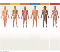
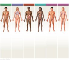
The human body contains 11 major organ systems, each with specific organs and functions. These systems work together to maintain homeostasis and overall health.
Organ System | Major Organs | Main Functions |
|---|---|---|
Integumentary | Skin, hair, sweat glands, nails | Protection, temperature regulation, sensory information |
Skeletal | Bones, cartilage, ligaments, bone marrow | Support, protection, mineral storage, blood cell formation |
Muscular | Skeletal muscles, tendons | Movement, support, heat generation |
Nervous | Brain, spinal cord, nerves, sense organs | Immediate response to stimuli, coordination, sensory input |
Endocrine | Pituitary, thyroid, adrenal glands, pancreas, gonads | Hormone production, long-term regulation, metabolism |
Cardiovascular | Heart, blood, blood vessels | Transport of nutrients, gases, wastes, heat distribution |
Lymphatic | Spleen, thymus, lymph nodes, vessels, tonsils | Defense against infection, return of tissue fluids |
Respiratory | Nasal cavities, lungs, trachea, bronchi | Gas exchange, sound production |
Digestive | Teeth, tongue, stomach, intestines, liver, pancreas | Food processing, nutrient absorption, water conservation |
Urinary | Kidneys, ureters, bladder, urethra | Waste excretion, water and ion balance, pH regulation |
Reproductive (Male/Female) | Testes, penis, ovaries, uterus, mammary glands, etc. | Production of sex cells and hormones, support of offspring |
Medical and Anatomical Terminology
Medical Terminology
Medical terms are constructed from word roots, prefixes, suffixes, and combining forms, often derived from Latin or Greek. Understanding these components aids in deciphering complex terms (e.g., histology = study of tissues).
Singular vs. Plural: Example: villus (singular), villi (plural); cortex (singular), cortices (plural).
Eponyms: Many commemorative names have been replaced by precise anatomical terms.
Anatomical Position and Landmarks
The anatomical position is the standard reference: standing, feet forward, arms at sides, palms forward. Other positions include supine (lying face up) and prone (lying face down).
Surface Anatomy: Identifies external features and landmarks for orientation.
Directional Terms
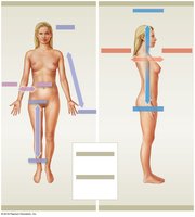
Directional terms describe locations and relationships of body parts:
Superior/Inferior: Above/below
Anterior (Ventral)/Posterior (Dorsal): Front/back
Medial/Lateral: Toward/away from midline
Proximal/Distal: Closer to/farther from point of attachment
Superficial/Deep: Near/far from body surface
Cranial/Caudal: Toward head/tail
Sectional Anatomy and Planes
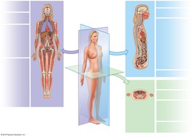
Sectional anatomy uses slices (sections) to visualize internal structures. Main planes include:
Frontal (Coronal) Plane: Divides body into anterior and posterior portions.
Sagittal Plane: Divides body into right and left portions (midsagittal = equal halves; parasagittal = unequal).
Transverse (Horizontal) Plane: Divides body into superior and inferior portions.
Body Cavities
Major Body Cavities and Their Functions
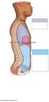
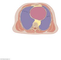
Body cavities protect internal organs and allow for changes in organ size and shape. The trunk is divided by the diaphragm into:
Thoracic Cavity: Contains pleural cavities (lungs) and pericardial cavity (heart), separated by the mediastinum.
Abdominopelvic Cavity: Contains abdominal (digestive organs) and pelvic (reproductive organs, bladder, rectum) cavities.
Serous Membranes
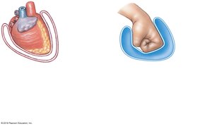
Serous membranes (serosa) line body cavities and cover organs, consisting of:
Parietal Layer: Lines the cavity wall.
Visceral Layer: Covers the organ.
Homeostasis
Definition and Importance
Homeostasis is the maintenance of a stable internal environment. All body systems contribute to keeping variables (e.g., temperature, fluid balance) within normal ranges.
Autoregulation: Local, automatic adjustment by cells, tissues, or organs.
Extrinsic Regulation: Regulation by nervous or endocrine systems.
Homeostatic Regulatory Mechanisms
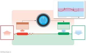
Consist of three main components:
Receptor: Detects changes (stimulus).
Control Center: Processes information and sends commands.
Effector: Carries out the response to restore balance.
Negative and Positive Feedback
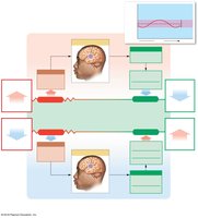
Negative Feedback: The effector's response negates the stimulus, maintaining homeostasis (e.g., body temperature regulation).
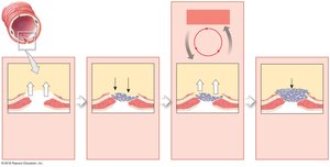
Positive Feedback: The response amplifies the original stimulus, moving the body away from homeostasis temporarily (e.g., blood clotting, childbirth).
Systems Integration and Disease
Organ systems work together to maintain dynamic equilibrium. Failure of homeostatic mechanisms leads to disease.
