 뒤로
뒤로Introduction to Anatomy and Physiology: Foundations, Homeostasis, and Imaging
스터디 가이드 - 스마트 노트
 자료에 맞춘 맞춤형 노트, 핵심 정의, 예시, 맥락을 확장해 제공합니다.
Introduction to Anatomy and Physiology
Importance of Anatomy and Physiology
Anatomy and Physiology (A&P) are foundational sciences in understanding the structure and function of the human body. Mastery of these subjects is essential for making informed decisions about health and for understanding disease processes.
Anatomy: The study of the structure of body parts and their relationships to one another.
Physiology: The study of the function of the body’s structural machinery.
Knowledge of A&P is crucial for healthcare professionals and anyone interested in understanding how the body works in both health and disease.
Historical Context and Methods
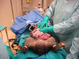
Anatomy is one of the oldest medical sciences, with roots in ancient dissections and vivisections. Early anatomists used animal models such as pigs and monkeys to study body structures.
Dissection and prosection are key methods for learning anatomy.
Modern anatomy education often uses prosection (dissection by an expert for teaching).

Key Concepts in Anatomy and Physiology
Definitions and Relationship
Anatomy and physiology are complementary disciplines. Anatomy focuses on structure, while physiology focuses on function. Both are necessary for a complete understanding of the human body.
Gross (Macroscopic) Anatomy: Study of large, visible structures.
Microscopic Anatomy: Study of structures too small to be seen with the naked eye (e.g., cytology and histology).
Physiology includes cell, organ, systemic, and clinical (pathological) physiology.
Levels of Structural Organization
The human body is organized in a hierarchy from simplest to most complex:
Atoms → Molecules → Organelles → Cells → Tissues → Organs → Organ Systems → Organism
Basic Life Characteristics
All living things share five basic characteristics:
Cellularity: Composed of one or more cells.
Metabolism: Obtain and use energy (anabolic and catabolic processes).
Reproduction: Ability to produce offspring (sexually or asexually).
Responsiveness: Ability to respond to environmental changes (homeostasis).
Adaptation and Evolution: Ability to adapt and evolve over generations.
Homeostasis
Definition and Importance
Homeostasis is the maintenance of a stable internal environment despite external changes. It is essential for normal physiological function and survival.
Involves negative and positive feedback loops.
Disruption of homeostasis can lead to disease or death.
Mechanisms of Homeostatic Regulation
Autoregulation (Intrinsic): Local, automatic adjustment by cells, tissues, or organs.
Extrinsic Regulation: Controlled by nervous and endocrine systems.
Components of Homeostatic Control
Receptor: Detects changes (stimuli).
Control Center: Processes information and determines response.
Effector: Carries out the response to restore balance.
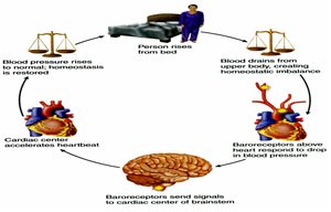
Negative Feedback Loops
Negative feedback opposes the initial change, helping to maintain homeostasis. Examples include regulation of body temperature, blood pressure, blood glucose, and calcium levels.
Body is brought back to a set point or normal range.
Positive Feedback Loops
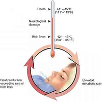
Positive feedback amplifies the initial stimulus, moving the system away from homeostasis. It is typically used for processes that need to be completed quickly, such as blood clotting, childbirth, and fever.
Can be dangerous if not controlled (e.g., high fever).
Anatomical Terminology
Body Positions and Directions
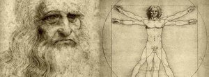
Standardized anatomical terms are used to describe body positions, directions, and planes.
Anatomical Position: Standing erect, facing forward, arms at sides, palms forward.
Supine: Lying on the back, face up.
Prone: Lying on the belly, face down.
Sectional Planes
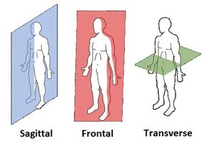
Sectional anatomy uses planes to describe slices of the body:
Sagittal (Median) Plane: Divides body into left and right.
Frontal (Coronal) Plane: Divides body into anterior and posterior.
Transverse (Horizontal) Plane: Divides body into superior and inferior.
Body Cavities and Membranes
Major Body Cavities
The body contains dorsal and ventral cavities, which house and protect internal organs.
Dorsal Cavity: Cranial and spinal cavities (brain and spinal cord).
Ventral Cavity: Thoracic and abdominopelvic cavities (heart, lungs, digestive organs, etc.).
Serous Membranes
Serous membranes line body cavities and cover organs, reducing friction and allowing movement.
Parietal Layer: Lines cavity walls.
Visceral Layer: Covers organs.
Medical Imaging Techniques
Overview of Imaging Modalities
Medical imaging is essential for visualizing internal structures and diagnosing disease. Each modality has specific uses and advantages.
X-rays
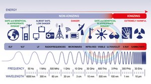
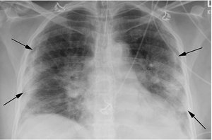
Uses high-energy radiation to visualize dense structures (e.g., bones).
Radiopaque structures appear white; radiolucent structures appear dark.
Computed Tomography (CT)
Combines X-ray images from multiple angles to create cross-sectional views.
Excellent for visualizing bone, soft tissue, and blood vessels.

Magnetic Resonance Imaging (MRI)
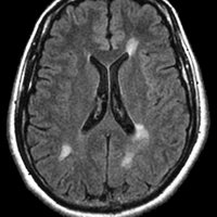
Uses magnetic fields and radio waves to produce detailed images of soft tissues.
Preferred for brain, spinal cord, and soft tissue injuries.
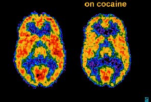
Nuclear Medicine and PET Scans
Use radioactive tracers to assess metabolic and physiological activity.
PET scans are valuable in oncology and neurology.
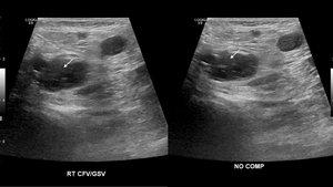
Ultrasound
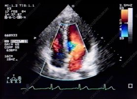
Uses high-frequency sound waves to create images of soft tissues and blood flow.
Commonly used in obstetrics, cardiology, and vascular studies.
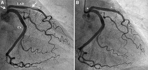
Digital Subtraction Angiography (DSA)
Invasive imaging technique for visualizing blood vessels using contrast dye and X-rays.
Used to assess vascular diseases and guide interventions.
Summary Table: Comparison of Imaging Modalities
Modality | Main Use | Advantages | Limitations |
|---|---|---|---|
X-ray | Bones, chest, teeth | Quick, inexpensive | Poor soft tissue contrast |
CT | Bone, soft tissue, blood vessels | Cross-sectional, detailed | Radiation exposure |
MRI | Brain, spinal cord, soft tissue | No radiation, high contrast | Expensive, time-consuming |
Ultrasound | Pregnancy, heart, vessels | Safe, real-time | Operator-dependent, limited by bone/air |
PET/Nuclear | Metabolic activity, cancer | Functional imaging | Radiotracer required |
Conclusion
Understanding the foundational concepts of anatomy and physiology, including homeostasis and medical imaging, is essential for students and professionals in the health sciences. Mastery of these topics provides the basis for further study in all areas of medicine and biology.
