 뒤로
뒤로Introduction to Anatomy & Physiology: Key Concepts, Body Systems, and Anatomical Organization
스터디 가이드 - 스마트 노트
 자료에 맞춘 맞춤형 노트, 핵심 정의, 예시, 맥락을 확장해 제공합니다.
Introduction to Anatomy & Physiology
Definition and Scope
Anatomy is the study of the structure of body parts and their relationships to one another, while physiology is the study of the function of the body’s structural machinery. These two disciplines are closely related, as structure determines function and vice versa.
Anatomy: Focuses on what body parts are called, where they are located, and how they are organized.
Physiology: Explains how anatomical structures work, including the chemical and physical processes involved.
Example: The heart’s anatomy (chambers, valves) explains its function (pumping blood).
Branches of Anatomy
Gross (Macroscopic) Anatomy: Study of large, visible structures. Includes:
Surface (Superficial) Anatomy: Study of external features.
Regional Anatomy: Study of specific areas (e.g., head, neck).
Systemic Anatomy: Study of organ systems (e.g., cardiovascular system).
Developmental Anatomy: Study of structural changes from conception to adulthood.
Pathological Anatomy: Study of structural changes caused by disease.
Microscopic Anatomy: Study of structures too small to be seen with the naked eye.
Cytology: Study of cells.
Histology: Study of tissues.
Branches of Physiology
Cell Physiology: Processes within and between cells.
Organ Physiology: Functions of specific organs (e.g., heart, stomach).
Systemic Physiology: Functions of organ systems (e.g., cardiovascular, nervous).
Pathological Physiology: Effects of diseases on organ or system function.
Levels of Organization in the Human Body
Hierarchy from Simple to Complex
The human body is organized into a hierarchy of structural levels, each building on the previous one:
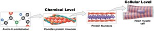
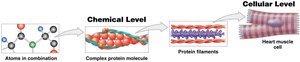
Chemical Level: Atoms combine to form molecules and compounds.
Cellular Level: Molecules combine to form cells, the basic units of life.
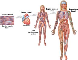
Tissue Level: Groups of similar cells working together to perform a function.
Organ Level: Two or more tissue types combine to form organs with specific functions.
Organ System Level: Groups of organs working together to perform complex functions.
Organism Level: All organ systems working together to maintain life and health.
Homeostasis and Feedback Mechanisms
Definition and Importance
Homeostasis is the body’s ability to maintain a stable internal environment despite changes in external conditions. It is essential for survival and proper function.
Examples: Regulation of body temperature, blood glucose, pH, water balance, and ion concentrations.
Multiple organ systems work together to maintain homeostasis.
The Three Components of Homeostatic Regulation
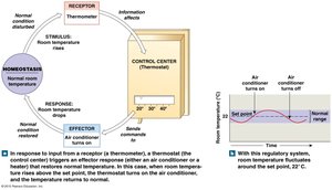
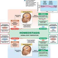
Receptor (Sensor): Detects changes in the environment (stimuli).
Control Center: Processes information and determines the appropriate response (often the brain or spinal cord).
Effector: Carries out the response to restore homeostasis (e.g., muscles, glands).
Negative vs. Positive Feedback
Negative Feedback: The primary mechanism for maintaining homeostasis. The response reduces or eliminates the original stimulus, returning the system to its set point.
Examples: Regulation of body temperature, blood glucose, blood pressure.
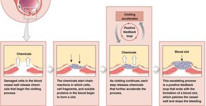
Positive Feedback: The response amplifies the original stimulus, moving the system further from its starting state. Used for processes that need rapid completion.
Examples: Blood clotting, labor contractions.
Systems Integration and Equilibrium
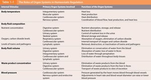
Multiple organ systems interact to maintain homeostasis, a state of dynamic equilibrium. Failure to maintain homeostasis can result in disease or death.
Internal Variable | Primary Organ Systems Involved | Functions of the Organ Systems |
|---|---|---|
Body temperature | Integumentary, Muscular, Cardiovascular, Nervous | Heat loss, heat production, heat distribution, coordination of heat loss and production |
Body fluid composition | Digestive, Cardiovascular, Urinary, Skeletal, Respiratory | Nutrient absorption, distribution, and release; nutrient distribution; control of nutrient loss; mineral storage and release; absorption of oxygen, elimination of carbon dioxide |
Body fluid volume | Urinary, Digestive, Integumentary, Cardiovascular, Lymphatic | Elimination or conservation of water; absorption of water; loss of water through perspiration; distribution of water; return of tissue fluid to blood |
Waste product concentration | Urinary, Cardiovascular, Digestive | Elimination of waste products from blood; transport of waste products to sites of excretion; elimination of waste products by the liver |
Blood pressure | Cardiovascular, Nervous, Endocrine | Heart and blood vessel actions; adjustment of heart rate and blood vessel diameter; adjustment of blood volume |
The 11 Organ Systems of the Human Body
Overview and Key Functions
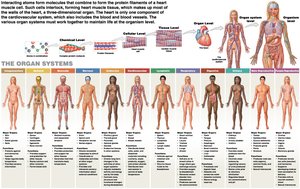
The human body is organized into 11 major organ systems, each with specific organs and functions essential for maintaining homeostasis and supporting life.
System | Major Organs | Primary Functions |
|---|---|---|
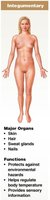
Integumentary | Skin, hair, sweat glands, nails | Protection, temperature regulation, sensory information |
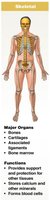
Skeletal | Bones, cartilage, ligaments, bone marrow | Support, protection, mineral storage, blood cell formation |
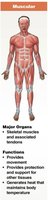
Muscular | Skeletal muscles, tendons | Movement, protection, heat generation |
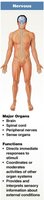
Nervous | Brain, spinal cord, nerves, sense organs | Immediate response, coordination, sensory information |
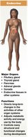
Endocrine | Pituitary, thyroid, pancreas, adrenal glands, gonads | Long-term changes, metabolic activity, development |
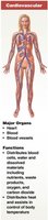
Cardiovascular | Heart, blood, blood vessels | Transport of cells and dissolved materials, heat distribution |
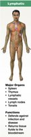
Lymphatic | Spleen, thymus, lymphatic vessels, lymph nodes, tonsils | Defense, returns tissue fluids to bloodstream |
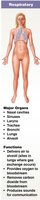
Respiratory | Nasal cavities, sinuses, larynx, trachea, bronchi, lungs, alveoli | Gas exchange, sound production |
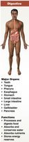
Digestive | Teeth, tongue, pharynx, esophagus, stomach, intestines, liver, gallbladder, pancreas | Digestion, absorption, water conservation, energy storage |
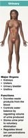
Urinary | Kidneys, ureters, urinary bladder, urethra | Excretion, water balance, pH regulation |
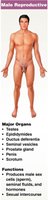
Reproductive (Male) | Testes, epididymides, ductus deferentia, seminal vesicles, prostate, penis, scrotum | Production of sperm, hormones, sexual intercourse |
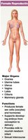
Reproductive (Female) | Ovaries, uterine tubes, uterus, vagina, labia, clitoris, mammary glands | Production of oocytes, hormones, supports embryo, milk production, sexual intercourse |
Body Cavities and Serous Membranes
Major Body Cavities
Body cavities are internal spaces that house organs and allow for changes in organ size and shape. They also protect organs from shock and damage.
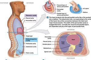
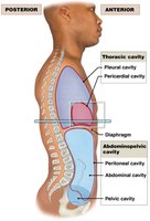
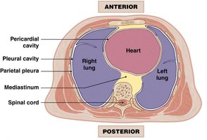
Thoracic Cavity: Superior cavity containing the lungs, heart, thymus, and parts of the esophagus and trachea.
Abdominopelvic Cavity: Inferior cavity containing digestive, urinary, and reproductive organs. Subdivided into abdominal and pelvic cavities.
The diaphragm separates the thoracic and abdominopelvic cavities.
Thoracic Cavity Subdivisions
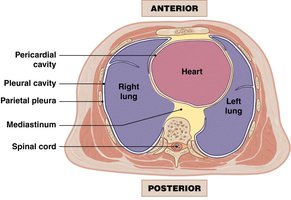
Pleural Cavities: Each houses a lung.
Mediastinum: Central compartment containing the heart (in the pericardial cavity), great vessels, trachea, esophagus, and thymus.
Serous Membranes
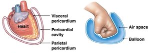
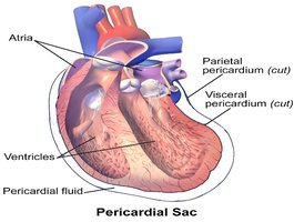
Serous membranes are double-layered membranes that line body cavities and cover organs. They secrete serous fluid to reduce friction.
Parietal Layer: Lines the cavity wall.
Visceral Layer: Covers the organ surface.
Examples:
Pericardium: Surrounds the heart.
Pleura: Surrounds the lungs.
Peritoneum: Surrounds abdominal organs.
Anatomical Terminology and Landmarks
Importance of Anatomical Terminology
Standardized anatomical terminology allows healthcare professionals to communicate clearly and precisely about body locations, regions, and movements.
Anatomical Position: Body standing upright, facing forward, arms at sides, palms forward.
Supine: Lying face up.
Prone: Lying face down.
Anatomical Regions and Quadrants
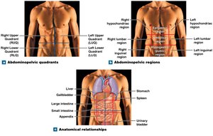
Abdominopelvic Quadrants: Right upper, left upper, right lower, left lower.
Abdominopelvic Regions: Nine regions for more precise localization (e.g., epigastric, umbilical, hypogastric).
Sectional Anatomy (Planes of the Body)
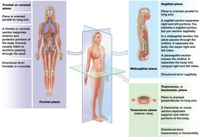
Frontal (Coronal) Plane: Divides body into anterior and posterior parts.
Sagittal Plane: Divides body into right and left parts (midsagittal = equal halves).
Transverse (Horizontal) Plane: Divides body into superior and inferior parts.
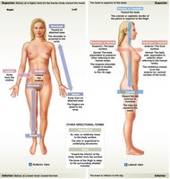
Anatomical Directions
Superior (Cranial): Toward the head.
Inferior (Caudal): Toward the feet.
Anterior (Ventral): Toward the front.
Posterior (Dorsal): Toward the back.
Medial: Toward the midline.
Lateral: Away from the midline.
Proximal: Closer to the point of attachment.
Distal: Farther from the point of attachment.
Superficial: Toward the surface.
Deep: Away from the surface.
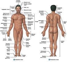
Common Anatomical Landmarks
Carpal: Wrist
Lumbar: Lower back
Thoracic: Chest
Gluteal: Buttock
Pedal: Foot
Brachial: Arm
Pollex: Thumb
Additional info: Mastery of these foundational concepts is essential for success in all subsequent topics in Anatomy & Physiology, including the study of tissues, organ systems, and clinical applications.
