 뒤로
뒤로Introduction to Anatomy & Physiology Lab: Microscopy, Anatomical Terminology, and the Cell
스터디 가이드 - 스마트 노트
 자료에 맞춘 맞춤형 노트, 핵심 정의, 예시, 맥락을 확장해 제공합니다.
Lab Safety and Responsibilities
Key Lab Safety Practices
Proper safety procedures are essential in the anatomy and physiology laboratory to prevent accidents and ensure a productive learning environment.
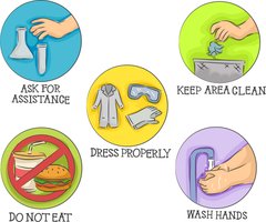
Dress Properly: Wear closed-toed shoes, lab coats, and safety goggles as required.
No Food or Drink: Eating or drinking in the lab is prohibited to avoid contamination and accidents.
Keep Area Clean: Clean your workspace and return all equipment to its proper place after use.
Ask for Assistance: Seek help from the instructor when handling unfamiliar equipment or chemicals.
Wash Hands: Always wash your hands after completing lab work.
Microscopy in Anatomy & Physiology
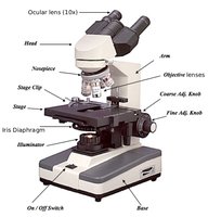
Parts of the Compound Light Microscope
Understanding the parts and functions of the microscope is foundational for studying cells and tissues in anatomy and physiology.
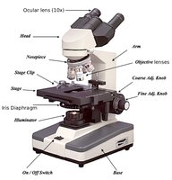
Ocular Lens (10x): The eyepiece through which you view the specimen.
Objective Lenses: Provide different magnification powers (commonly 4x, 10x, 40x; 100x oil immersion is not used in this course).
Stage: Platform where the slide is placed.
Coarse and Fine Focus Knobs: Used to bring the specimen into focus.
Illuminator and Iris Diaphragm: Control the amount and focus of light on the specimen.
Objective Lenses and Magnification
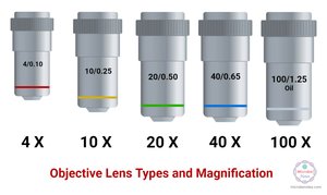
Microscopes use a combination of ocular and objective lenses to magnify specimens. The total magnification is calculated by multiplying the ocular lens power by the objective lens power.
Common Objective Lenses: 4x (scanning), 10x (low power), 40x (high power), 100x (oil immersion; not used in this lab).
Total Magnification Formula:
Example: Using a 40x objective lens: total magnification.
Example: Using a 4x objective lens: total magnification.
Field of View
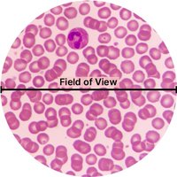
The field of view is the visible area when looking through the microscope. It is largest at the lowest magnification and decreases as magnification increases.
Scanning Power (4x): Largest field of view.
High Power (40x): Smaller field of view, but greater detail.
Basic Steps for Using a Microscope
Proper technique ensures clear visualization and prevents damage to the microscope or slides.
Turn on the microscope and adjust the light.
Lower the stage using the coarse focus knob.
Start with the scanning (4x) objective lens.
Place the slide on the stage and center the specimen over the light.
Use the coarse focus knob to bring the specimen into view, then fine focus for clarity.
Switch to higher magnification objectives as needed, using only the fine focus knob.
When finished, return to the lowest objective and lower the stage.
Anatomical Terminology and Body Orientation
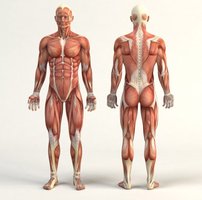
Anatomical Position
The anatomical position is a standardized reference for describing locations and directions on the human body. The body stands upright, facing forward, with arms at the sides and palms facing forward.
Relative Anatomical Terminology
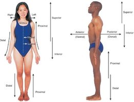
Relative terms describe the location of one body part in relation to another.
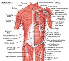
Superficial: Closer to the body surface.
Deep: Farther from the body surface.
Anterior (Ventral): Toward the front of the body.
Posterior (Dorsal): Toward the back of the body.
Superior: Toward the head or upper part of a structure.
Inferior: Away from the head or toward the lower part of a structure.
Medial: Toward the midline of the body.
Lateral: Away from the midline of the body.
Proximal: Closer to the point of attachment or origin.
Distal: Farther from the point of attachment or origin.
Body Planes and Sections
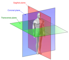
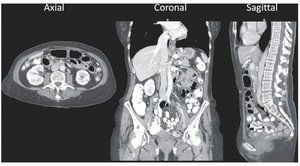
Body planes are imaginary lines used to divide the body into sections for anatomical study and medical imaging.
Sagittal Plane: Divides the body into right and left parts. If equal, it is called midsagittal; otherwise, parasagittal.
Frontal (Coronal) Plane: Divides the body into anterior and posterior (front and back) parts.
Transverse (Axial) Plane: Divides the body into superior (upper) and inferior (lower) parts.
Absolute Anatomical Terminology
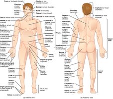
Absolute terms refer to specific regions of the body, often using Latin or Greek roots. The adjective form is commonly used in clinical and anatomical descriptions.
Nasal (nose), Oral (mouth), Cervical (neck), Brachial (arm), Femoral (thigh), etc.
Example: The term "nasal region" is used more often than "nasus" (the noun form).
The Cell: Structure and Function
Introduction to Cells
The cell is the smallest unit of all life. Cells vary in structure and function, and not all cells contain every organelle.
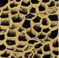
First Cells Observed: Dead cork cells, which appeared empty under early microscopes.
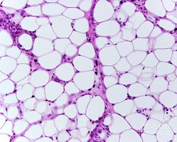
Cell Diversity: Examples include plant cells, animal cells, and specialized cells such as adipose (fat) cells.
Animal Cell Structure
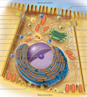
Animal cells contain various organelles, each with specialized functions. Not every cell has every structure, and organelles can differ between cell types.
Key Organelles: Nucleus, mitochondria, endoplasmic reticulum, Golgi apparatus, lysosomes, microvilli, vacuoles.
Specialized Cells: Some, like skeletal muscle cells, are multinucleate.
Tissues: Cells are organized into tissues, which form organs.
The Cell Cycle and Cellular Division
Phases of the Cell Cycle
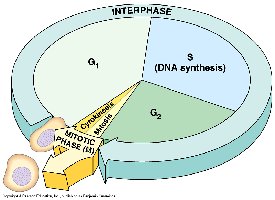
Cells undergo a life cycle that includes periods of growth, DNA replication, and division. Most of a cell's life is spent in interphase, which is subdivided into G1, S, and G2 phases. Checkpoints regulate progression through the cycle.
Interphase: Cell grows (G1), replicates DNA (S), and prepares for division (G2).
Mitosis: Division of the nucleus, followed by cytokinesis (division of the cell membrane).
Purpose and Regulation of Cell Division
Mitotic cell division is essential for growth, development, and tissue repair in multicellular organisms. The rate of cell division varies by cell type.
Rapidly Dividing Cells: Intestinal lining cells divide every few days.
Rarely Dividing Cells: Heart muscle cells may divide only once or twice in a lifetime.
Meiosis: In gonads, cells divide by meiosis to produce gametes (sperm or eggs).

Mitosis: Stages and Importance
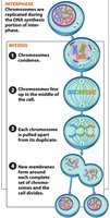
Mitosis produces two genetically identical daughter cells for growth or replacement. Large numbers of cells are replaced daily by mitosis (e.g., skin, fat, and blood cells).
Stages of Mitosis: Prophase, Metaphase, Anaphase, Telophase, followed by Cytokinesis (often abbreviated as iPMATC).
Clinical Relevance: Loss of cell cycle control can lead to cancer (uncontrolled mitosis).
Microscopy and Identification of Mitosis Stages
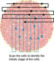
In laboratory practice, students should be able to identify the stages of mitosis in prepared slides, such as those of whitefish blastula cells.
Lab Activity: Identify interphase, prophase, metaphase, anaphase, telophase, and cytokinesis under the microscope.
Summary Table: Key Concepts in Lab 1
Topic | Key Points | Example/Application |
|---|---|---|
Lab Safety | Dress code, no food/drink, clean area, ask for help | Wear goggles, wash hands after lab |
Microscopy | Parts, magnification, field of view, focusing steps | Calculate total magnification: |
Anatomical Terminology | Relative and absolute terms, body planes | Anterior vs. posterior, sagittal plane |
Cell Structure | Organelles, cell diversity, tissue formation | Identify nucleus, mitochondria, etc. on models |
Cell Cycle & Mitosis | Interphase, mitosis stages, checkpoints | Observe mitosis in whitefish slides |
