 뒤로
뒤로Introduction to Anatomy & Physiology: Organization, Terminology, and Homeostasis
스터디 가이드 - 스마트 노트
 자료에 맞춘 맞춤형 노트, 핵심 정의, 예시, 맥락을 확장해 제공합니다.
Introduction to Anatomy & Physiology
Anatomy and Physiology Compared
Anatomy is the study of the structure and form of the body, while physiology focuses on how the body functions. These disciplines are closely connected, as understanding structure aids in understanding function and vice versa.
Microscopic Anatomy: Examines structures not visible to the naked eye, typically using a microscope.
Gross (Macroscopic) Anatomy: Studies structures visible to the naked eye, often through dissection.
Characteristics of Living Things
Organization: All organisms exhibit a complex structure and order.
Metabolism: The sum of all chemical reactions in the body.
Anabolism: Building larger molecules from smaller ones.
Catabolism: Breaking down large molecules into smaller ones.
Growth and Development: Organisms grow and develop by assimilating materials from their environment.
Responsiveness: The ability to sense and react to stimuli.
Regulation/Adaptation: Adjusting internal functions to accommodate environmental changes; includes homeostasis.
Reproduction: Producing new cells for growth, maintenance, and repair; with gametes, can produce new organisms.
Levels of Organization in the Human Body
From Simplest to Most Complex
Chemical Level: Atoms, molecules, macromolecules, and organelles.
Cellular Level: Cells are the basic units of structure and function.
Tissue Level: Groups of similar cells performing common functions.
Epithelial Tissue: Covers surfaces and lines cavities.
Connective Tissue: Supports, protects, and binds organs.
Muscle Tissue: Produces movement.
Nervous Tissue: Conducts nerve impulses.
Organ Level: Two or more tissue types working together for specific functions.
Organ System Level: Related organs working together for a common function.
Organismal Level: All body systems functioning together in an individual.
Overview of Organ Systems
Integumentary System: Skin, hair, nails; protects body, regulates temperature.
Skeletal System: Bones, cartilage; supports, protects, stores minerals, forms blood cells.
Muscular System: Muscles; movement, posture, heat production.
Nervous System: Brain, spinal cord, nerves; controls and coordinates body activities.
Endocrine System: Glands; hormone production, regulation of metabolism, growth, and reproduction.
Cardiovascular System: Heart, blood vessels; transports nutrients, gases, wastes.
Lymphatic System: Lymph nodes, vessels, spleen; immune defense, fluid balance.
Respiratory System: Lungs, airways; gas exchange.
Urinary System: Kidneys, bladder; removes waste, regulates water and electrolytes.
Digestive System: Stomach, intestines; breaks down food, absorbs nutrients, eliminates waste.
Male Reproductive System: Testes, ducts; produces sperm and hormones.
Female Reproductive System: Ovaries, uterus; produces eggs, supports pregnancy.
Anatomic Position and Terminology
Anatomic Position
The standard reference position for anatomical terminology: upright stance, feet parallel and flat, arms at sides, palms facing forward, head level, eyes forward.
Sections and Planes
Coronal (Frontal) Plane: Divides body into anterior (front) and posterior (back) parts.
Transverse (Cross-sectional) Plane: Divides body into superior (upper) and inferior (lower) parts.
Midsagittal (Median) Plane: Divides body into equal left and right halves.
Sagittal Plane: Parallel to midsagittal, divides into unequal left and right parts.
Oblique Plane: Passes through at an angle.
Directional Terms
Anterior (Ventral): Toward the front
Posterior (Dorsal): Toward the back
Superior: Toward the head
Inferior: Toward the feet
Cranial: Toward the head
Caudal: Toward the tail
Rostral: Toward the nose/mouth
Medial: Toward the midline
Lateral: Away from the midline
Deep: Further from the surface
Superficial: Closer to the surface
Proximal: Closer to point of attachment
Distal: Farther from point of attachment
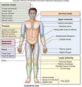
Regional Anatomy
Axial Region: Head, neck, trunk, and spine (central axis of the body).
Appendicular Region: Limbs and their attachments (arms, legs, shoulders, hips).
Body Cavities and Membranes
Major Body Cavities
Posterior Aspect: Cranial cavity (houses brain), vertebral canal (houses spinal cord); both encased in bone.
Ventral Cavity: Larger, anterior; includes thoracic and abdominopelvic cavities, not fully encased in bone.
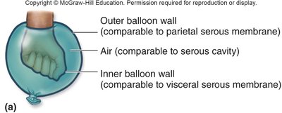
Serous Membranes
Parietal Layer: Lines internal surface of body wall.
Visceral Layer: Covers external surface of organs.
Serous Cavity: Space between layers, filled with serous fluid to reduce friction.
Thoracic Cavity
Mediastinum: Central compartment; contains heart, thymus, esophagus, trachea, major blood vessels.
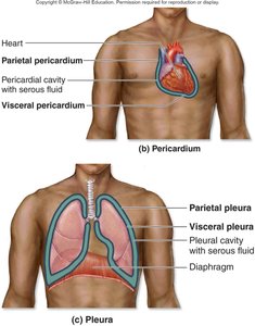
Pericardium: Two-layered serous membrane around the heart.
Parietal Pericardium: Outer layer, forms sac around heart.
Visceral Pericardium: Covers heart's surface.
Pericardial Cavity: Space with serous fluid between layers.
Pleura: Two-layered serous membrane around lungs.
Parietal Pleura: Lines thoracic wall.
Visceral Pleura: Covers lungs.
Pleural Cavity: Space with serous fluid between layers.
Abdominopelvic Cavity
Abdominal Cavity: Superior; contains digestive organs, kidneys, most of ureters.
Pelvic Cavity: Inferior; contains distal large intestine, bladder, reproductive organs.
Peritoneum: Two-layered serous membrane lining abdominopelvic cavity.
Parietal Peritoneum: Lines internal walls.
Visceral Peritoneum: Covers external surfaces of organs.
Peritoneal Cavity: Space with serous fluid between layers.
Abdominopelvic Regions and Quadrants
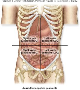
The abdominopelvic cavity is divided for clinical and anatomical reference:
9 Regions: Epigastric, umbilical, hypogastric, right/left hypochondriac, right/left lumbar, right/left iliac.
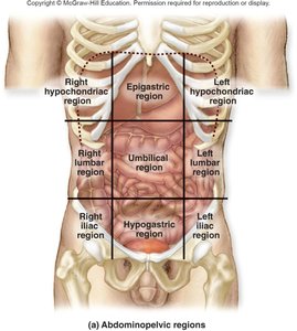
4 Quadrants: Right upper (RUQ), left upper (LUQ), right lower (RLQ), left lower (LLQ).
Homeostasis: Maintaining Internal Stability
Definition and Importance
Homeostasis is the ability of an organism to maintain a stable internal environment despite changes in external or internal conditions. It is essential for survival and proper function.
Components of Homeostatic Systems
Receptor: Detects changes in a variable (e.g., temperature sensors in skin).
Control Center: Interprets input and initiates response (often the brain or endocrine gland).
Effector: Carries out the response to restore balance (e.g., muscles, glands).
Stimulus: Change in a regulated variable.
Receptor detects the change.
Receptor sends information to control center.
Control center processes and sends output to effector.
Effector acts to restore homeostasis.
Negative Feedback
Most homeostatic processes use negative feedback.
The response moves the variable in the opposite direction of the stimulus to maintain a normal range.
Example: Body temperature regulation.
Positive Feedback
Less common; the response reinforces the stimulus until a climactic event occurs, then homeostasis is restored.
Example: Blood clotting, childbirth contractions.
Clinical View: Normal Ranges
Body Temperature: 98.6°F
Blood Glucose: 80–110 mg/dL
Blood Pressure: 90–120/60–80 mm Hg
Additional info: Homeostatic imbalance can lead to disease or dysfunction, highlighting the importance of these regulatory mechanisms in health and medicine.
