 뒤로
뒤로Introduction to Anatomy & Physiology: Orientation and Fundamentals
스터디 가이드 - 스마트 노트
 자료에 맞춘 맞춤형 노트, 핵심 정의, 예시, 맥락을 확장해 제공합니다.
Introduction to Anatomy & Physiology
Definitions and Importance
Anatomy and physiology are foundational sciences for understanding the structure and function of the human body. Anatomy is the study of the structure of body parts and their relationships to one another, while physiology is the study of the function of the body and its parts. Studying these subjects is essential for health professionals to diagnose, treat, and understand human health and disease.
Levels of Biological Organization
Hierarchy of Life
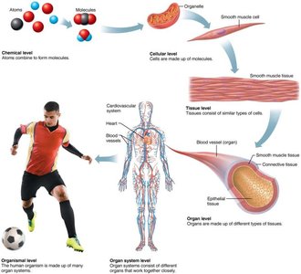
The human body is organized into a hierarchy of structural levels, each building upon the previous:
Chemical Level: Atoms combine to form molecules and macromolecules.
Cellular Level: Cells are the basic structural and functional units of life.
Tissue Level: Groups of similar cells form tissues.
Organ Level: Organs are made up of different types of tissues.
Organ System Level: Organ systems consist of different organs working together.
Organismal Level: The human organism is made up of many organ systems functioning together.
Organ Systems Overview
Major Organ Systems and Functions
The body is composed of several organ systems, each with specific components and functions:
Integumentary System: Skin; protection.
Skeletal System: Bones; protection, support, hemopoiesis.
Muscular System: Muscles; movement, body temperature regulation.
Nervous System: Brain, spinal cord, nerves; communication and control.
Endocrine System: Glands; control and regulation via hormones.
Cardiovascular System: Heart, blood vessels; transportation of nutrients and wastes.
Lymphatic System: Spleen, lymph nodes; immune protection, fluid return.
Respiratory System: Lungs, airways; air distribution and gas exchange.
Digestive System: GI tract, accessory organs; nutrient breakdown and absorption.
Urinary System: Kidneys, bladder; filter blood and excrete wastes.
Reproductive System: Gonads; produce gametes and hormones.
Homeostasis
Definition and Regulation
Homeostasis is the body's ability to maintain a stable internal environment despite external changes. When homeostasis is not maintained, disease or dysfunction can occur. Regulation can be intrinsic (local, automatic response) or extrinsic (involving nervous or endocrine systems).
Feedback Mechanisms
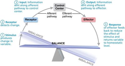
Homeostasis is regulated by feedback mechanisms, which consist of three main components:
Receptor: Detects changes (stimulus).
Control Center: Receives input, processes information, determines set point, sends commands.
Effector: Responds to commands and brings about change, either opposing or enhancing the stimulus.
Negative Feedback
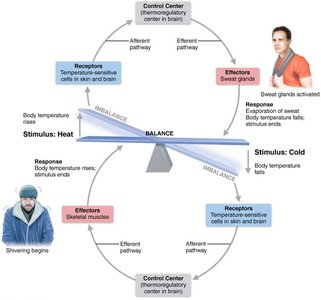
Negative feedback mechanisms reduce the effect of the original stimulus, returning the variable to its set point. This is the most common type of feedback in the body. Examples include regulation of blood glucose, body temperature, and blood calcium.
Positive Feedback
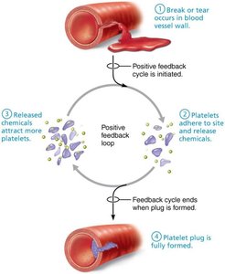
Positive feedback mechanisms enhance the original stimulus, leading to an amplified response. These are less common and typically occur in situations requiring a rapid outcome, such as blood clotting, uterine contractions during childbirth, and breastfeeding.
Anatomical Terminology
Standard Anatomical Position (SAP)
The Standard Anatomical Position is a common reference position used in anatomy: standing upright, facing forward, arms at sides, palms facing forward, feet together.
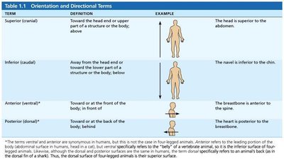
Directional Terms
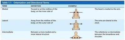
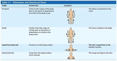
Directional terms are used to describe the locations of structures relative to other structures or locations in the body. These terms are essential for clear communication in anatomy.
Term | Definition | Example |
|---|---|---|
Superior (cranial) | Toward the head or upper part of a structure | The head is superior to the abdomen. |
Inferior (caudal) | Away from the head end or toward the lower part | The navel is inferior to the chin. |
Anterior (ventral) | Toward or at the front of the body | The breastbone is anterior to the spine. |
Posterior (dorsal) | Toward or at the back of the body | The heart is posterior to the breastbone. |
Term | Definition | Example |
|---|---|---|
Medial | Toward the midline of the body | The heart is medial to the arm. |
Lateral | Away from the midline of the body | The arms are lateral to the chest. |
Intermediate | Between a more medial and a more lateral structure | The collarbone is intermediate between the breastbone and shoulder. |
Term | Definition | Example |
|---|---|---|
Proximal | Closer to the origin of the body part | The elbow is proximal to the wrist. |
Distal | Farther from the origin of the body part | The knee is distal to the thigh. |
Superficial (external) | Toward or at the body surface | The skin is superficial to the skeletal muscles. |
Deep (internal) | Away from the body surface | The lungs are deep to the skin. |
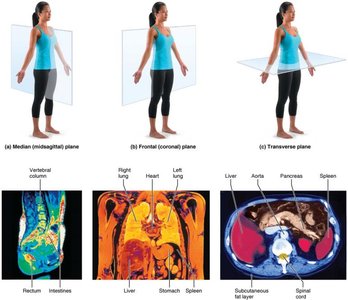
Body Planes
Body planes are imaginary lines used to divide the body into sections:
Transverse (horizontal) plane: Divides the body into upper and lower portions.
Frontal (coronal) plane: Divides the body into front and back portions.
Sagittal plane: Divides the body into right and left parts.
Midsagittal plane: Divides the body into equal right and left halves.
Oblique plane: Divides the body at an angle.
Body Cavities
Major Body Cavities
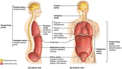
Internal organs are housed within enclosed spaces called body cavities. The two major body cavities are:
Dorsal Body Cavity: Contains the cranial cavity (brain) and vertebral cavity (spinal cord).
Ventral Body Cavity: Contains the thoracic cavity (heart, lungs) and abdominopelvic cavity (digestive, urinary, reproductive organs).
Serous Membranes
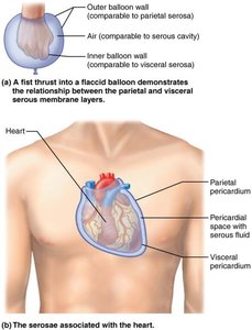
The subdivisions of the ventral body cavity are lined with serous membranes, which have two layers:
Parietal layer: Lines the internal surface of the body wall.
Visceral layer: Covers the external surface of organs (viscera).
Serous cavity: Fluid-filled space between layers, containing serous fluid to reduce friction.
Examples include the pericardium (heart), pleurae (lungs), and peritoneum (abdominal organs).
Abdominopelvic Quadrants and Regions
Quadrants
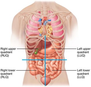
The abdominopelvic cavity is divided into four quadrants for clinical reference:
Right Upper Quadrant (RUQ)
Left Upper Quadrant (LUQ)
Right Lower Quadrant (RLQ)
Left Lower Quadrant (LLQ)
Regions
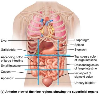
For more detailed localization, the abdominopelvic cavity is divided into nine regions:
Right hypochondriac
Epigastric
Left hypochondriac
Right lumbar
Umbilical
Left lumbar
Right iliac (inguinal)
Hypogastric (pubic)
Left iliac (inguinal)
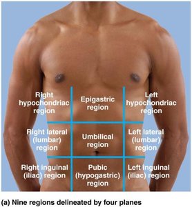
Clinical Application
These divisions help clinicians describe the location of pain, injuries, or pathology. For example, the appendix is found in the right lower quadrant (RLQ) and the right iliac region.
Summary Table: Levels of Organization
Level | Description | Example |
|---|---|---|
Chemical | Atoms and molecules | Water, proteins |
Cellular | Cells | Muscle cell |
Tissue | Groups of cells | Muscle tissue |
Organ | Groups of tissues | Heart |
Organ System | Groups of organs | Cardiovascular system |
Organism | All organ systems | Human body |
Key Equations
Homeostatic Feedback Loop (Generalized)
The feedback loop can be represented as:
Additional info:
Some context and definitions were inferred for completeness.
Tables were recreated and expanded for clarity.
