 뒤로
뒤로Introduction to Anatomy & Physiology: Structure, Function, and Organization of the Human Body
스터디 가이드 - 스마트 노트
 자료에 맞춘 맞춤형 노트, 핵심 정의, 예시, 맥락을 확장해 제공합니다.
Introduction to Anatomy & Physiology
Overview of Anatomy and Physiology
Anatomy and physiology are foundational disciplines in the health sciences, focusing on the structure and function of the human body. Understanding anatomical terminology is essential for accurate communication among health professionals.
Anatomy: The study of body structure, including the organization and relationships of body parts.
Physiology: The study of how body parts function and interact to sustain life.
Principle of Complementarity: Structure and function are inseparable; what a structure can do depends on its form.
Branches of Anatomy and Physiology
Various branches of anatomy and physiology allow for specialized study of the human body.
Branch | Focus |
|---|---|
Developmental Biology | Growth and development from fertilization to death |
Embryology | First eight weeks after fertilization |
Cell Biology | Cellular structure and function |
Histology | Microscopic structure of tissues |
Gross Anatomy | Structures visible without a microscope |
Systemic Anatomy | Structure of specific body systems |
Regional Anatomy | Specific regions of the body |
Imaging Anatomy | Internal structures visualized by imaging techniques |
Clinical Anatomy | Application to medical practice |
Pathological Anatomy | Structural changes associated with disease |
Branch of Physiology | Focus |
|---|---|
Molecular Physiology | Functions of molecules like proteins and DNA |
Neurophysiology | Functional properties of nerve cells |
Endocrinology | Hormones and their regulatory functions |
Cardiovascular Physiology | Heart and blood vessel functions |
Immunology | Body's defense mechanisms |
Respiratory Physiology | Functions of air passageways and lungs |
Renal Physiology | Kidney functions |
Exercise Physiology | Cell and organ changes due to muscular activity |
Pathophysiology | Functional changes due to disease and aging |
Levels of Structural Organization
Hierarchy of Organization
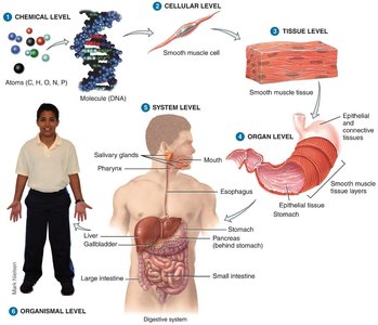
The human body is organized from the smallest chemical level to the whole organism level. Each level builds upon the previous, creating complex structures and functions.
Chemical Level: Atoms, molecules, and organelles
Cellular Level: Single cells
Tissue Level: Groups of similar cells
Organ Level: Two or more types of tissues
Organ System Level: Organs working together
Organismal Level: All organ systems combined
Major Organ Systems of the Human Body
Overview of Organ Systems
The human body contains eleven major organ systems, each with specific components and functions that contribute to homeostasis and overall health.
Integumentary System: Skin, hair, nails; protects body, regulates temperature, detects sensations
Skeletal System: Bones, joints, cartilages; supports and protects body, aids movement, stores minerals
Muscular System: Skeletal muscles; movement, posture, heat production
Nervous System: Brain, spinal cord, nerves, sense organs; regulates activities, interprets changes, responds
Endocrine System: Hormone-producing glands; regulates activities via hormones
Cardiovascular System: Heart, blood vessels; transports nutrients, regulates temperature, defends against disease
Lymphatic/Immune System: Lymphatic fluid, vessels, spleen, thymus, lymph nodes; returns proteins, carries lipids, immune responses
Respiratory System: Lungs, air passageways; gas exchange, regulates acid-base balance
Digestive System: GI tract, accessory organs; breakdown and absorption of food, eliminates wastes
Urinary System: Kidneys, ureters, bladder, urethra; eliminates wastes, regulates blood composition
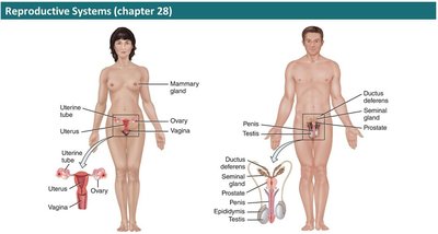
Reproductive System: Gonads, associated organs; produces gametes, hormones, supports reproduction
Basic Life Processes and Homeostasis
Characteristics of Life
All living things share certain characteristics that distinguish them from nonliving things. These processes are essential for maintaining life and homeostasis.
Metabolism: All chemical reactions in the body
Responsiveness: Ability to detect and respond to changes
Movement: Motion of the body, organs, cells
Growth: Increase in size and complexity
Differentiation: Development of specialized cells
Reproduction: Formation of new cells or organisms
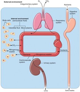
Homeostasis: Maintenance of stable internal conditions
Homeostasis and Feedback Mechanisms
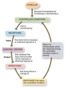
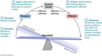
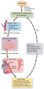
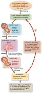
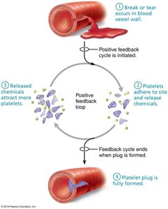
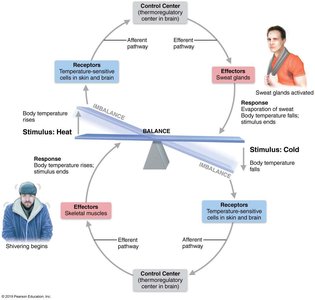
Homeostasis is regulated by feedback loops, which can be negative or positive. These mechanisms ensure the body's internal environment remains stable despite external changes.
Negative Feedback: Reverses a change to restore balance (e.g., temperature regulation)
Positive Feedback: Enhances a change until a specific outcome is achieved (e.g., labor contractions)
Anatomical Terminology
Body Positions
Standardized anatomical positions allow for consistent reference and communication.
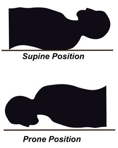
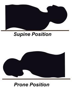
Anatomical Position: Standing erect, facing forward, arms at sides, palms forward, feet flat
Prone Position: Lying face down
Supine Position: Lying face up
Regional Names
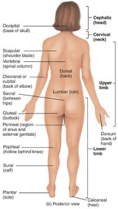
Regional terms designate specific areas within body divisions for reference.
Axial: Head, neck, trunk
Appendicular: Limbs (arms and legs)
Directional Terms
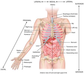
Directional terms are used to precisely locate one part of the body relative to another.
Term | Definition | Example |
|---|---|---|
Superior | Toward the head | The heart is superior to the liver |
Inferior | Away from the head | The stomach is inferior to the lungs |
Anterior (Ventral) | Nearer to the front | The sternum is anterior to the heart |
Posterior (Dorsal) | Nearer to the back | The esophagus is posterior to the trachea |
Medial | Nearer to the midline | The ulna is medial to the radius |
Lateral | Farther from the midline | The lungs are lateral to the heart |
Proximal | Nearer to limb attachment | The humerus is proximal to the radius |
Distal | Farther from limb attachment | The phalanges are distal to the carpals |
Superficial | Toward the surface | The ribs are superficial to the lungs |
Deep | Away from the surface | The ribs are deep to the skin |
Planes and Sections
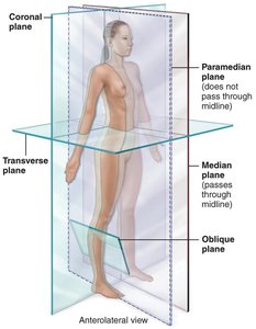
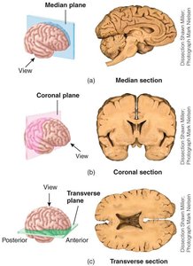
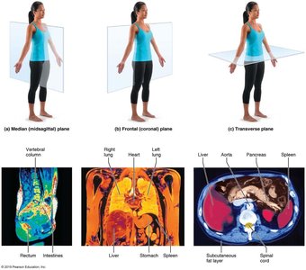
Planes are imaginary lines along which the body or organs may be cut for anatomical study. Sections are named after the plane.
Sagittal Plane: Divides body into right and left parts
Midsagittal Plane: Divides body exactly at midline
Parasagittal Plane: Off-center sagittal division
Frontal (Coronal) Plane: Divides body into anterior and posterior parts
Transverse (Horizontal) Plane: Divides body into superior and inferior parts
Oblique Section: Cuts at an angle other than 90°
Body Cavities and Membranes
Body Cavities
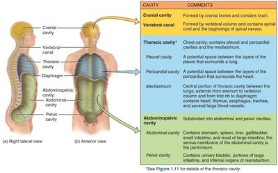
Body cavities are spaces within the body that protect, separate, and support internal organs.
Dorsal Cavity: Cranial and vertebral cavities
Ventral Cavity: Thoracic and abdominopelvic cavities, separated by the diaphragm
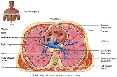
Thoracic Cavity: Pleural cavities (lungs), pericardial cavity (heart), mediastinum
Abdominopelvic Cavity: Abdominal (digestive organs) and pelvic (bladder, reproductive organs, rectum) cavities
Serous Membranes
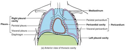
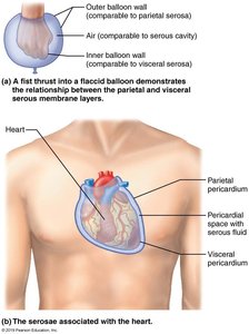
Serous membranes are thin, double-layered membranes that cover the viscera within the thoracic and abdominal cavities and line the walls of these cavities.
Visceral Layer: Covers the organ
Parietal Layer: Lines the cavity wall
Pericardium: Covers the heart
Pleura: Covers the lungs
Peritoneum: Covers abdominal organs
Retroperitoneal: Organs posterior to the peritoneum
Abdominopelvic Regions and Quadrants
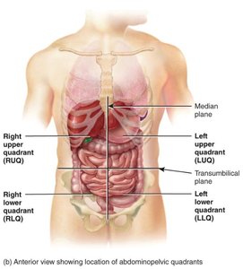
The abdominopelvic cavity can be divided into regions or quadrants to describe the location of organs.
Quadrants: Right upper (RUQ), left upper (LUQ), right lower (RLQ), left lower (LLQ)
Regions: Right hypochondriac, epigastric, left hypochondriac, right lumbar, umbilical, left lumbar, right iliac, hypogastric, left iliac
Medical Imaging Procedures
Overview of Medical Imaging
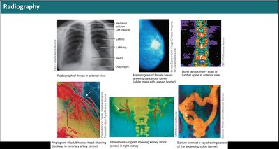
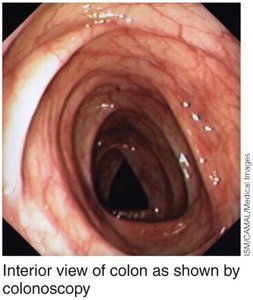
Medical imaging techniques allow physicians to view internal structures for diagnosis and treatment.
X-ray (Radiograph): Uses x-rays to produce images of internal structures
MRI: Uses magnetic fields and radio waves to create detailed images
CT Scan: Uses x-ray beams to produce cross-sectional images
Ultrasound: Uses high-frequency sound waves to produce images
Endoscopy: Uses a lighted instrument to view internal organs
CCTA: Uses contrast medium and x-rays for coronary vessel imaging
PET Scan: Uses positron-emitting substances to show tissue activity
Radionuclide Scanning: Uses radioactive substances to show tissue activity
Aging and Homeostasis
Aging Process
Aging is characterized by a progressive decline in the body's ability to restore homeostasis, impacting all body systems and increasing vulnerability to stress and disease.
Structural and functional changes occur with age
Homeostatic imbalance increases risk of disease
Sample Questions for Review
At what levels of structural organization would an exercise physiologist study the human body?
What quadrant is the appendix located in? The liver?
The ankle is _______ to the knee. Distal/proximal/superior/inferior
What are the two major regions of the body?
Why is the extracellular fluid called the internal environment of the body?
Additional info: This study guide covers the essential concepts from Chapter 1 of Anatomy & Physiology, including terminology, organization, homeostasis, and medical imaging, with expanded academic context for clarity and completeness.
