 뒤로
뒤로Introduction to Anatomy & Physiology: Structure, Function, and Tissue Types
스터디 가이드 - 스마트 노트
 자료에 맞춘 맞춤형 노트, 핵심 정의, 예시, 맥락을 확장해 제공합니다.
Human Anatomy & Physiology
Definition and Scope
Anatomy and Physiology (A & P) are foundational sciences that study the structure and function of the human body. Understanding both is essential for recognizing how injury and disease affect normal biological processes.
Anatomy: The study of the structure of body parts. It includes both gross anatomy (large, visible structures) and microscopic anatomy (structures requiring magnification).
Physiology: The study of the functions of body parts and how they work together.
Relationship: Structure and function are closely linked; healthy structures enable normal function, while disease or injury disrupts both.
Gross Anatomy
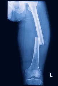
Studies large body parts visible to the naked eye, such as the heart, brain, teeth, and bones.
Example: A broken femur disrupts normal movement and causes pain.
Microscopic Anatomy
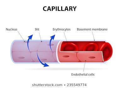
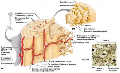
Requires a microscope to study smaller structures, such as capillaries, nerves, taste buds, cancer cells, and Haversian canals.
Terms for Anatomical Position, Body Planes, and Body Cavities
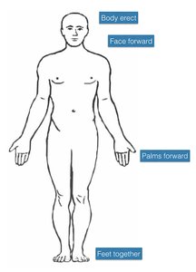
Anatomical Position
The standard reference position for anatomical terminology: standing erect, facing forward, arms at the sides, and palms facing forward.
Body Planes
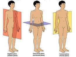
Body planes are imaginary lines used to divide the body for study and description.
Sagittal Plane: Divides the body into right and left halves.
Transverse Plane: Divides the body into superior (top) and inferior (bottom) halves.
Frontal (Coronal) Plane: Divides the body into anterior (front) and posterior (back) parts.
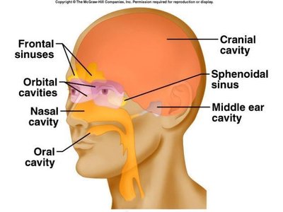
Body Cavities
Body cavities are spaces within the body that house organs and protect them.
Dorsal Body Cavity: Posterior cavity, includes cranial (brain) and spinal (spinal cord) cavities.
Ventral Body Cavity: Anterior cavity, divided by the diaphragm into thoracic (heart, lungs) and abdominopelvic (digestive, urinary, reproductive organs) cavities.
Directional Terms
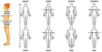
Directional terms describe the locations of structures relative to other structures or locations in the body.
Superior (cranial): Closer to the head
Inferior (caudal): Away from the head
Anterior (ventral): Toward the front
Posterior (dorsal): Toward the back
Medial: Toward the midline
Lateral: Away from the midline
Proximal: Closer to the trunk
Distal: Away from the trunk
Superficial: Toward the surface
Deep: Away from the surface
Human Body Systems
Overview of Body Systems
The human body consists of 12 major systems, each composed of organs working together to perform essential functions.
Integumentary System: Skin layers; protects, eliminates waste, regulates temperature.
Skeletal System: Bones, cartilage, ligaments; supports, protects, produces blood cells.
Muscular System: Cardiac, smooth, skeletal muscle; movement, heart pumping, digestion.
Nervous System: Brain, spinal cord, nerves; sensory input, movement, cognition.
Special Sensory System: Eyes, ears, smell, taste organs; vision, hearing, smell, taste.
Endocrine System: Glands; hormone secretion.
Respiratory System: Lungs, airways; oxygen delivery, CO2 removal.
Cardiovascular System: Heart, blood vessels; transport of oxygen, nutrients, waste.
Lymphatic System: Lymph vessels, nodes; fluid return, immunity.
Digestive System: Esophagus, stomach, intestines; nutrient breakdown and absorption.
Urinary System: Kidneys, bladder; waste removal.
Reproductive System: Testes, ovaries, uterus; offspring production.
Homeostasis
Definition and Mechanisms
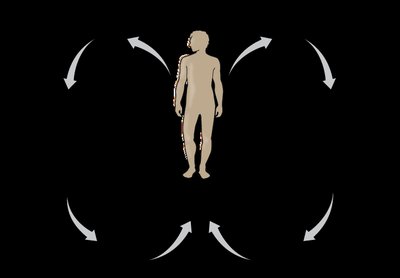
Homeostasis is the internal balance maintained by the body. It is achieved through coordinated actions of all organ systems, especially the nervous and endocrine systems.
Negative Feedback: Reduces a disruptive influence to restore balance. Example: If body temperature rises above 37℃, the hypothalamus triggers sweating and blood vessel dilation to cool the body.
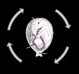
Positive Feedback: Increases a disruptive influence until a specific end point is reached. Example: Uterine contractions during childbirth increase until delivery occurs.
Homeostatic Imbalance: Occurs when the body cannot maintain homeostasis, leading to illness or death.
Tissues
Overview and Classification
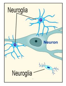
Tissues are groups of cells working together for a common function. Histology is the study of tissues. There are four main types: nervous, muscle, connective, and epithelial.
Nervous Tissue
Concentrated in the brain and spinal cord, with nerves extending throughout the body.
Transmits information via electrical signals.
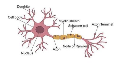
Composed of neurons (nerve cells) and neuroglia (supporting cells).
Muscle Tissue
Skeletal Muscle: Striated, voluntary, attached to skeleton, causes movement and facial expressions.
Cardiac Muscle: Striated, involuntary, found only in the heart, contracts to pump blood.
Smooth Muscle: Non-striated, involuntary, found in walls of hollow organs (blood vessels, GI tract, bladder).
Epithelial Tissue
Covers body surfaces and lines cavities.
Functions: protection, control of substance entry/exit, sensory information, secretion.
Attached to underlying connective tissue at the basement membrane.
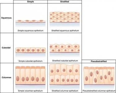
Cell shapes: squamous (flat), cuboidal (square), columnar (rectangular).
Arrangements: simple (single layer), stratified (multiple layers), pseudostratified (appears layered).
Types of Epithelial Tissue
Simple | Stratified | Pseudostratified | |
|---|---|---|---|
Squamous | Simple squamous epithelium | Stratified squamous epithelium | |
Cuboidal | Simple cuboidal epithelium | Stratified cuboidal epithelium | |
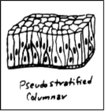
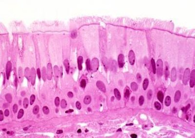
Columnar | Simple columnar epithelium | Stratified columnar epithelium | Pseudostratified columnar epithelium |
Simple Squamous: Diffusion and filtration; found in air sacs of lungs and capillaries.
Simple Cuboidal: Secretion and absorption; found in kidney tubules, ducts, ovaries.
Stratified Cuboidal: Lining and protection; found in ducts of sweat, mammary, salivary glands.
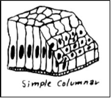
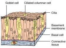
Simple Columnar: Secretion and absorption; found in digestive tract and uterus; contains goblet cells and microvilli.
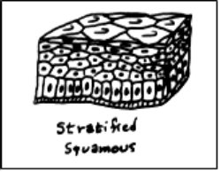
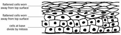
Stratified Squamous: Protection; found in skin and mouth.
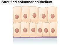
Stratified Columnar: Secretion and protection; rare, found in male urethra, conjunctiva, ducts.
Pseudostratified Columnar: Secretion and cilia-aided movement; found in air passages and reproductive tubes.
Connective Tissue
Most abundant tissue; binds, protects, supports, fills space, stores fat.
Types: loose (areolar, reticular, adipose), dense (tendons, ligaments), cartilage (hyaline, elastic, fibrocartilage), bone, blood.
Loose Connective Tissue
Areolar: Binds organs to skin and each other; forms thin membranes.
Adipose: Stores fat.
Reticular: Provides framework in lymph nodes, bone marrow, spleen.
Dense Connective Tissue
Tendons: Connect muscle to bone.
Ligaments: Connect bone to bone; slow healing due to poor blood supply.
Cartilage
Hyaline: Most common; found on ends of bones, ribs, nose, trachea, larynx.
Elastic: Flexible; found in outer ear and epiglottis.
Fibrocartilage: Strongest; found between vertebrae, knee, pubic joint.
Bone (Osseous Tissue)
Protects organs, supports body, provides framework for muscle movement.
Blood
Transports, regulates, protects; carries oxygen, nutrients, wastes, and CO2.
Additional info: Academic context was added to clarify tissue functions and anatomical terminology.
