 뒤로
뒤로Introduction to Anatomy and Physiology: The Human Body—An Orientation
스터디 가이드 - 스마트 노트
 자료에 맞춘 맞춤형 노트, 핵심 정의, 예시, 맥락을 확장해 제공합니다.
Introduction to Anatomy and Physiology
Overview of the Chapter
This chapter introduces the foundational concepts of anatomy and physiology, providing a framework for understanding the human body. It explores the relationship between structure and function, the organization of the body, requirements for life, homeostasis, and the terminology used to describe anatomical structures and regions.
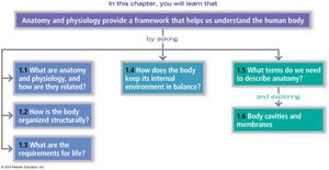
What are Anatomy and Physiology, and How Are They Related?
Definitions and Complementarity
Anatomy is the study of the structure of body parts and their relationships to one another.
Physiology is the study of the function of body parts—how they work to carry out life-sustaining activities.
Topics of anatomy include gross (macroscopic) anatomy (structures visible to the naked eye) and microscopic anatomy (structures too small to be seen without magnification).
Physiology is often subdivided by organ systems and focuses on cellular and molecular events.
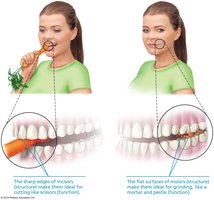
The principle of complementarity of structure and function states that what a structure can do depends on its specific form.
Example: The sharp edges of incisors are ideal for cutting food, while the flat surfaces of molars are suited for grinding.
How is the Body Organized Structurally?
Levels of Structural Organization
The human body is organized into a hierarchy of structural levels, each building on the previous one:
Chemical level: Atoms combine to form molecules.
Cellular level: Cells are made up of molecules and are the basic unit of life.
Tissue level: Tissues consist of groups of similar cells with a common function.
Organ level: Organs are made up of different types of tissues working together.
Organ system level: Organ systems consist of different organs that work closely together.
Organismal level: The human organism is made up of many organ systems working together to maintain life.
The Body’s Organ Systems and Their Major Functions
Overview of Organ Systems
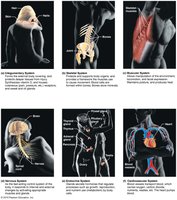
The body is composed of multiple organ systems, each with specific functions essential for survival:
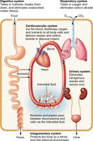
Integumentary System: Protects the body, regulates temperature, and provides sensory information.
Skeletal System: Supports and protects organs, provides a framework for muscles, and forms blood cells.
Muscular System: Allows movement, maintains posture, and produces heat.
Nervous System: Fast-acting control system that responds to internal and external changes.
Endocrine System: Glands secrete hormones that regulate processes such as growth, reproduction, and metabolism.
Cardiovascular System: Transports blood, nutrients, gases, and wastes.
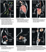
Lymphatic/Immune System: Returns leaked fluids to blood, disposes of debris, and houses immune cells.
Respiratory System: Supplies blood with oxygen and removes carbon dioxide.
Digestive System: Breaks down food, absorbs nutrients, and eliminates waste.
Urinary System: Eliminates nitrogenous wastes and regulates water, electrolyte, and acid-base balance.
Reproductive System: Produces offspring and sex hormones.
Interrelationships Among Body Organ Systems
Systemic Integration
Organ systems do not function in isolation; they interact to maintain the internal environment of the body. For example, the digestive, respiratory, cardiovascular, and urinary systems work together to provide nutrients, remove wastes, and maintain homeostasis.
What are the Requirements for Life?
Necessary Life Functions
Maintaining boundaries: Separation between internal and external environments (e.g., skin, cell membranes).
Movement: Includes movement of the body and substances within the body.
Responsiveness: Ability to sense and respond to stimuli.
Digestion: Breakdown of ingested food into simple molecules.
Metabolism: All chemical reactions within the body.
Excretion: Removal of wastes from the body.
Reproduction: Production of offspring and new cells.
Growth: Increase in size of a body part or the organism as a whole.
Survival Needs
Nutrients: Chemicals for energy and cell building.
Oxygen: Essential for energy production in cells.
Water: Most abundant substance in the body; provides environment for chemical reactions.
Normal body temperature: Necessary for proper metabolic reactions.
Appropriate atmospheric pressure: Required for adequate breathing and gas exchange.
Homeostasis: Maintaining Internal Balance
Definition and Mechanisms
Homeostasis is the ability of the body to maintain a relatively stable internal environment, despite changes in the external environment. It is maintained through complex communication systems involving receptors, control centers, and effectors.
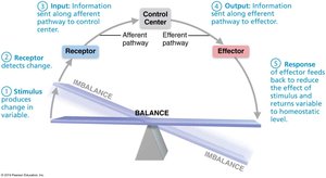
Stimulus: Produces change in a variable.
Receptor: Detects change and sends information to the control center.
Control Center: Determines set point, analyzes input, and coordinates response.
Effector: Carries out the response to restore balance.
Feedback: Response feeds back to reduce or amplify the effect of the stimulus.
Negative and Positive Feedback Mechanisms
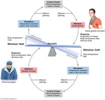
Negative feedback: Most homeostatic mechanisms; reduce or shut off the original stimulus (e.g., regulation of body temperature).
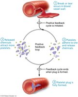
Positive feedback: Amplifies the original stimulus; used for processes that need a definitive end point (e.g., blood clotting).
Homeostatic imbalance can lead to disease or dysfunction.
Describing Anatomy: Terminology and Body Organization
Anatomical Position and Regional Terms
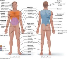
Anatomical position: Body erect, feet slightly apart, palms facing forward, thumbs pointing away from the body. Right and left refer to the subject's right and left.
The body is divided into axial (head, neck, trunk) and appendicular (limbs) regions.
Regional terms specify areas within these divisions.
Directional Terms
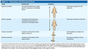
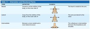
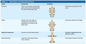
Directional terms are used to explain where one body part is in relation to another. Common terms include superior/inferior, anterior/posterior, medial/lateral, proximal/distal, superficial/deep.
Term | Definition | Example |
|---|---|---|
Superior (cranial) | Toward the head or upper part of a structure | The head is superior to the abdomen. |
Inferior (caudal) | Away from the head or toward the lower part of a structure | The navel is inferior to the chin. |
Anterior (ventral) | Toward or at the front of the body | The breastbone is anterior to the spine. |
Posterior (dorsal) | Toward or at the back of the body | The heart is posterior to the breastbone. |
Term | Definition | Example |
|---|---|---|
Medial | Toward or at the midline of the body | The heart is medial to the arm. |
Lateral | Away from the midline of the body | The arms are lateral to the chest. |
Intermediate | Between a more medial and a more lateral structure | The collarbone is intermediate between the breastbone and shoulder. |
Term | Definition | Example |
|---|---|---|
Proximal | Closer to the origin of the body part or point of attachment | The elbow is proximal to the wrist. |
Distal | Farther from the origin of a body part or point of attachment | The knee is distal to the thigh. |
Superficial (external) | Toward or at the body surface | The skin is superficial to the muscles. |
Deep (internal) | Away from the body surface; more internal | The lungs are deep to the skin. |
Body Planes and Sections
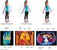
Sagittal plane: Divides the body into right and left parts. A midsagittal plane lies on the midline; a parasagittal plane is offset from the midline.
Frontal (coronal) plane: Divides the body into anterior and posterior parts.
Transverse (horizontal) plane: Divides the body into superior and inferior parts.
Body Cavities and Membranes
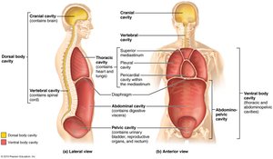
Dorsal body cavity: Protects the nervous system; includes cranial and vertebral cavities.
Ventral body cavity: Houses internal organs (viscera); includes thoracic and abdominopelvic cavities.
Thoracic cavity: Contains pleural cavities (lungs) and mediastinum (heart and other structures).
Abdominopelvic cavity: Contains abdominal (digestive organs) and pelvic (urinary, reproductive organs) cavities.
Serous Membranes
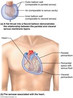
Serous membranes (serosa): Thin, double-layered membranes that cover the walls of the ventral body cavity and the outer surfaces of organs.
Parietal serosa: Lines cavity walls.
Visceral serosa: Covers organs within the cavity.
Serous fluid between layers reduces friction.
Named for the specific organs they cover (e.g., pericardium for heart, pleura for lungs, peritoneum for abdominal organs).
Abdominopelvic Regions and Quadrants
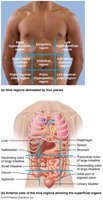
The abdominopelvic cavity is divided for clinical and anatomical reference into four quadrants or nine regions.
Quadrants: Right upper (RUQ), left upper (LUQ), right lower (RLQ), left lower (LLQ).
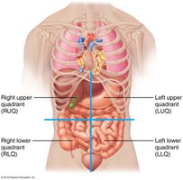
Regions: Right hypochondriac, epigastric, left hypochondriac, right lumbar, umbilical, left lumbar, right iliac (inguinal), hypogastric (pubic), left iliac (inguinal).
Additional info: Understanding these foundational concepts is essential for further study in anatomy and physiology, as they provide the language and framework for describing the human body in health and disease.
