 뒤로
뒤로Introduction to Anatomy: Organization, Terminology, and Microscopy
스터디 가이드 - 스마트 노트
 자료에 맞춘 맞춤형 노트, 핵심 정의, 예시, 맥락을 확장해 제공합니다.
Foundations of Anatomy
Levels of Organization
The human body is organized into several hierarchical levels, each with increasing complexity. Understanding these levels is fundamental to the study of anatomy and physiology.
Chemical Level: Atoms and molecules essential for life.
Cellular Level: Basic structural and functional units of life.
Tissue Level: Groups of similar cells performing a common function.
Organ Level: Structures composed of two or more tissue types working together.
Organ System Level: Groups of organs that perform related functions.
Organismal Level: The complete living being.
There are 11 major organ systems in the human body:
Integumentary
Skeletal
Muscular
Nervous
Endocrine
Cardiovascular
Lymphatic
Respiratory
Digestive
Urinary
Reproductive
Anatomical Position and Directional Terms
Anatomical Position
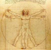
The anatomical position is a standardized stance used as a reference in describing the location and relation of body parts. The body stands upright, facing forward, with arms at the sides and palms facing forward. All directional terms are based on this position.
Directional Terms
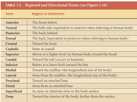
Directional terms are used to describe the locations of structures relative to other structures or locations in the body. They are always referenced from the anatomical position and the body's central mass or midline.
Term | Region or Reference |
|---|---|
Anterior | The front; before |
Ventral | The belly side (equivalent to anterior when referring to human body) |
Posterior | The back; behind |
Dorsal | The back (equivalent to posterior when referring to human body) |
Cranial | Toward the head |
Cephalic | Same as cranial |
Superior | Above; at a higher level (in human body, toward the head) |
Caudal | Toward the tail (coccyx in humans) |
Inferior | Below; at a lower level; toward the feet |
Medial | Toward the midline (the longitudinal axis of the body) |
Lateral | Away from the midline (the longitudinal axis of the body) |
Proximal | Toward an attached base |
Distal | Away from an attached base |
Superficial | At, near, or relatively close to the body surface |
Deep | Toward the interior of the body; farther from the surface |
Body Planes and Sections
Major Body Planes
Body planes are imaginary lines used to divide the body into sections for anatomical study and medical imaging.
Transverse (Horizontal) Plane: Divides the body into superior (upper) and inferior (lower) portions.
Coronal (Frontal) Plane: Divides the body into anterior (front) and posterior (back) portions.
Sagittal Plane: Divides the body into left and right portions. A midsagittal plane divides the body into equal left and right halves.
Abdominopelvic Quadrants
Clinical Relevance
The abdominopelvic cavity is divided into four quadrants for diagnostic and descriptive purposes. Common clinical conditions such as gallstones and appendicitis are often localized using these quadrants.
Body Cavities and Serous Membranes
Major Body Cavities
A body cavity is an enclosed space within the body that houses organs. Major cavities include the cranial, thoracic, abdominal, and pelvic cavities.
Serous membranes are protective sheets of connective tissue that line body cavities.
Visceral layer: The inner layer, directly covering the organ.
Parietal layer: The outer layer, lining the cavity wall.
Introduction to Microscopy
Microscope Parts and Function
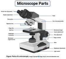
Microscopy is essential for studying cells and tissues in anatomy. The compound light microscope is the most common instrument used in laboratories.
Ocular lens: Usually 10x magnification.
Objective lenses: Typically 4x, 10x, 40x, and 100x (oil immersion).
Total magnification: Calculated by multiplying the ocular lens by the objective lens.
Example: Using a 4x objective lens, total magnification = 10 (ocular) x 4 (objective) = 40.
Microscopy Tips
Always start with the lowest magnification (4x).
Bring the stage up to its highest position, but avoid contact with the objective lens.
Use the coarse-focus knob to bring the specimen into view, then fine-focus for clarity.
When increasing magnification, lower the stage before switching objectives to avoid damaging the slide.
Never use the coarse-focus knob at high magnifications (40x or above).
As magnification increases, the field of view decreases.
The Metric System in Anatomy
Units and Conversions
The metric system is the standard for measurement in anatomy and the sciences. Students should be comfortable converting between kilometers, meters, centimeters, and millimeters in both directions.
1 kilometer (km) = 1,000 meters (m)
1 meter (m) = 100 centimeters (cm)
1 centimeter (cm) = 10 millimeters (mm)
Example: 2.5 cm = 25 mm
