뒤로
뒤로Introduction to Human Anatomy and Physiology: Chapter 1 Study Notes
스터디 가이드 - 스마트 노트
 자료에 맞춘 맞춤형 노트, 핵심 정의, 예시, 맥락을 확장해 제공합니다.
Introduction to Human Anatomy and Physiology
Overview of Anatomy and Physiology
Anatomy and physiology are foundational sciences in understanding the human body. Anatomy is the study of the physical structure of body parts and their relationships, while physiology focuses on how these structures function to sustain life. Both disciplines are closely linked, as structure determines function.
Gross Anatomy: Study of large, visible structures (regional, systemic, surface anatomy).
Microscopic Anatomy: Study of structures too small to be seen with the naked eye (histology, cytology).
Developmental Anatomy: Study of structural changes throughout the lifespan (e.g., embryology).
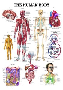
Physiology examines how anatomical structures work, often at the cellular or molecular level. Examples include renal physiology (kidney function), neurophysiology (nervous system function), and cardiovascular physiology (heart and blood function).
Relationship Between Structure and Function
The form of a body part determines its function. For example, the mineralization of bone provides strength, and heart valves prevent blood backflow. Slight anatomical variations exist among individuals, but extreme variations are rare.
Sex: Biological attributes based on chromosomes, gene expression, and hormones (e.g., reproductive anatomy).
Gender: Psychological and social constructs related to identity and expression.
Levels of Structural Organization
Hierarchy of Organization
The human body is organized into a hierarchy of structural levels, each building on the previous one:
Chemical Level: Atoms combine to form molecules, which make up organelles.
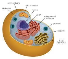
Cellular Level: Cells are the smallest living units, composed of organelles.
Tissue Level: Groups of similar cells form tissues with specific functions.
Organ Level: Organs are made of at least two tissue types and perform unique functions.
Organ System Level: Organs work together in systems to accomplish common purposes.
Organism Level: All systems function together to sustain the living human organism.
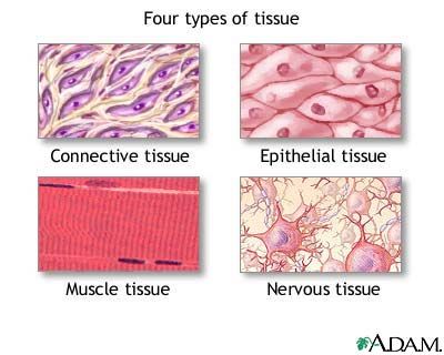
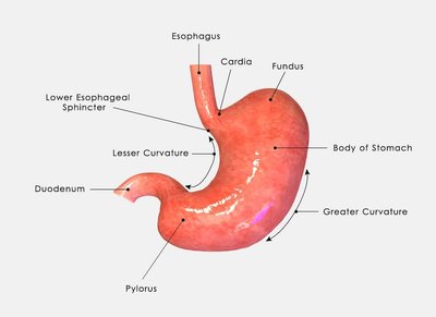
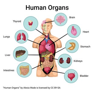
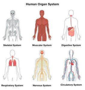
Necessary Life Functions
Key Life Functions
To maintain life, the human body must perform several essential functions:
Maintaining Boundaries: Separation of internal and external environments (e.g., plasma membrane, skin).
Movement: Movement of body parts, substances, and cells (muscle contractility).
Responsiveness: Ability to sense and respond to stimuli (e.g., reflexes, breathing rate).
Digestion: Breakdown of food into absorbable molecules for distribution via blood.
Metabolism: All chemical reactions in the body, including catabolism (breakdown) and anabolism (synthesis). Regulated by hormones.
Excretion: Removal of metabolic wastes (e.g., urea, CO2, feces).
Reproduction: Cellular division for growth/repair and production of offspring.
Growth: Increase in size of body parts or the organism as a whole.
Survival Needs
Essential Requirements for Survival
The body requires specific substances and conditions to survive. Imbalances can be harmful.
Nutrients: Carbohydrates (energy), proteins (cell building), fats (energy storage), vitamins and minerals (chemical reactions and structure).
Oxygen: Required for metabolic reactions; brain damage occurs within minutes without it.
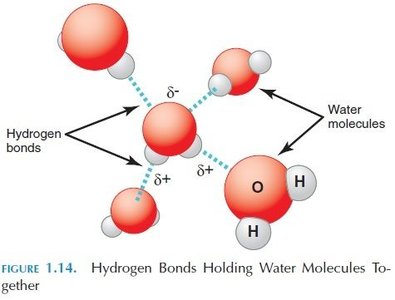
Water: Most abundant substance in the body; necessary for chemical reactions and as a medium for secretions/excretions.
Normal Body Temperature: Maintained around 37°C (98.6°F); deviations affect chemical reactions.
Atmospheric Pressure: Required for proper breathing and gas exchange in the lungs.
Homeostasis
Definition and Importance
Homeostasis is the maintenance of a stable internal environment despite external changes. It involves dynamic equilibrium, where variables are kept within narrow limits. All organ systems contribute to homeostasis.
Law of Mass Balance: Input must equal output to maintain constant levels of substances.
Homeostatic Control Mechanisms
Homeostasis is regulated mainly by the nervous and endocrine systems. Three components are involved:
Receptor: Detects changes (stimuli) and sends information to the control center.
Control Center: Determines the set point and appropriate response; sends output to the effector.
Effector: Carries out the response to restore balance (e.g., muscle contraction, gland secretion).
Types of Feedback
Negative Feedback: Reduces or shuts off the original stimulus, returning the variable to the set point. Most common type (e.g., body temperature regulation, blood glucose control).
Positive Feedback: Enhances the original stimulus, causing a greater deviation (e.g., labor contractions, blood clotting).
Feedforward Response: Anticipatory response before a change occurs (e.g., salivation before eating).
Homeostatic Imbalance
Disruption of homeostasis increases the risk of disease and is associated with aging. When feedback mechanisms are overwhelmed, negative feedback may fail, and positive feedback can become destructive.
Organization of Anatomy: Anatomical Terminology
Importance of Anatomical Terms
Standardized anatomical terminology is essential for accurately describing body locations, positions, and movements, reducing errors in healthcare communication.
Anatomical Position
The reference position for anatomical terminology: body erect, feet slightly apart, palms facing forward, thumbs pointing away from the body.
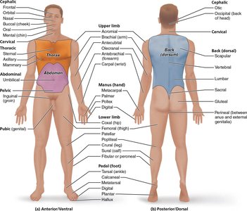
Directional Terms
Used to describe the location of one body part relative to another, always based on the anatomical position. Right and left refer to the subject's sides, not the observer's.
Body Planes
Sagittal Plane: Divides the body into left and right parts. Midsagittal is at the midline; parasagittal is offset.
Frontal (Coronal) Plane: Divides the body into anterior (front) and posterior (back) parts.
Transverse Plane: Divides the body into superior (upper) and inferior (lower) parts.
Oblique Section: A cut at an angle other than 90°; rarely used.
Body Cavities and Membranes
Major Body Cavities
Dorsal Body Cavity: Protects the nervous system; includes the cranial cavity (brain) and vertebral cavity (spinal cord), both covered by meninges.
Ventral Body Cavity: Houses internal organs (viscera); includes the thoracic and abdominopelvic cavities.
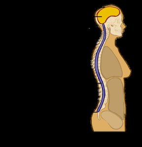
Ventral Body Cavity Subdivisions
Thoracic Cavity: Contains two pleural cavities (lungs), mediastinum (other thoracic organs), and pericardial cavity (heart).
Abdominopelvic Cavity: Separated from thoracic cavity by the diaphragm; includes abdominal cavity (digestive organs, spleen, liver) and pelvic cavity (bladder, reproductive organs, rectum).
Serous Membranes
Thin, double-layered membranes lining ventral body cavities and covering organs. They secrete serous fluid for lubrication.
Parietal Serosa: Lines cavity walls.
Visceral Serosa: Covers organs.
Pleura: Lungs (parietal pleurae, visceral pleurae).
Pericardium: Heart (parietal pericardium, visceral pericardium).
Peritoneum: Abdominopelvic cavity (parietal peritoneum, visceral peritoneum).
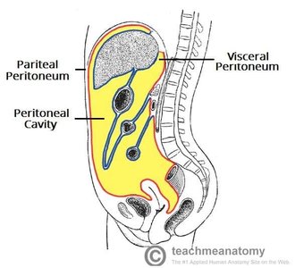
Clinical Correlation
Inflammation of serous membranes can cause pain and dysfunction:
Pleurisy: Inflammation of the pleura (lungs).
Peritonitis: Inflammation of the peritoneum (abdomen).
Abdominopelvic Regions and Quadrants
The abdominopelvic cavity is divided for clinical reference:
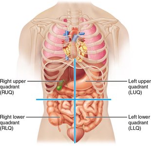
Four Quadrants: Right Upper (RUQ), Right Lower (RLQ), Left Upper (LUQ), Left Lower (LLQ).
Nine Regions: Used for more precise localization of organs and pain.