 뒤로
뒤로Introduction to Human Anatomy & Physiology: Organization, Subdivisions, and Homeostasis
스터디 가이드 - 스마트 노트
 자료에 맞춘 맞춤형 노트, 핵심 정의, 예시, 맥락을 확장해 제공합니다.
Anatomy: Subdivisions and Approaches
Definition and Overview
Anatomy is the scientific study of the structure and interrelationships of body parts. It provides the foundational knowledge necessary to understand how the human body is organized and how its parts relate to one another.
Gross Anatomy: Study of structures visible to the naked eye, without magnification.
Microscopic Anatomy: Study of structures requiring magnification, such as cells and tissues.
Developmental Anatomy: Study of structural changes from conception through old age.
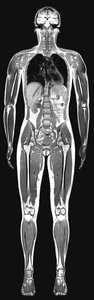
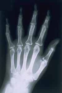
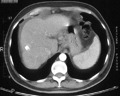
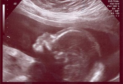
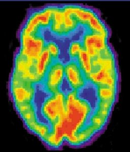
Radiographic Anatomy: Study of body structures using imaging techniques (e.g., X-ray, CT, MRI, PET, Ultrasound).
Pathological Anatomy: Study of structural changes associated with disease.
Gross Anatomy Subdivisions
Systemic Anatomy: Study of all structures within a particular body system (e.g., skeletal, muscular, nervous systems).
Regional Anatomy: Study of all structures within a specific region of the body (e.g., head, neck, thorax).
Surface Anatomy: Study of internal structures as they relate to the body surface.
Microscopic Anatomy
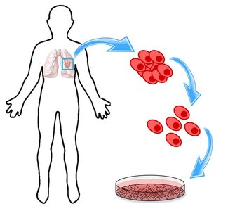
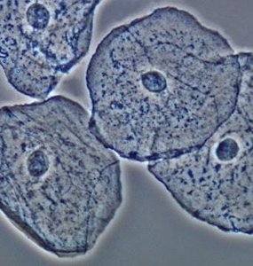
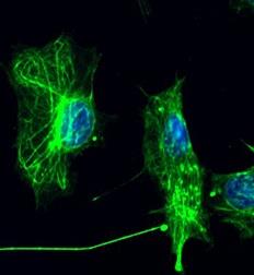
Cytology: Study of cells, the smallest units displaying characteristics of life.
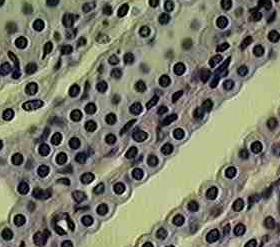
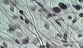
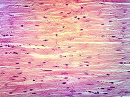
Histology: Study of tissues, which are groups of two or more cells with a common function.
There are four primary tissue classes in the body:
Epithelial tissue
Connective tissue
Muscle tissue
Nerve tissue
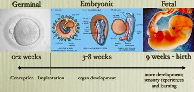
Developmental Anatomy
Developmental anatomy examines structural changes from conception through old age, including:
Embryology: Study of development from fertilization to the end of the eighth week.
Fetology: Study of development from the ninth week to birth.
Neonatology: Study of newborns.
Gerontology: Study of aging and the elderly.
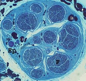

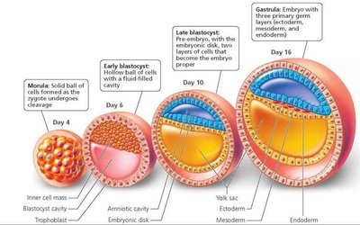
During early embryonic development, three primary germ layers are formed:
Ectoderm: Outer layer; forms nervous system and skin.
Mesoderm: Middle layer; forms muscle and connective tissues.
Endoderm: Inner layer; forms epithelial lining of digestive, respiratory, and urogenital systems and associated glands.

Radiographic Anatomy
Radiographic anatomy utilizes imaging techniques to visualize internal structures:
X-ray: Uses electromagnetic radiation to visualize solid structures.
Computed Tomography (CT): Motorized X-ray source circles the body to generate 3D images.
Positron Emission Tomography (PET): Uses radioactive tracers to highlight metabolically active tissues.
Magnetic Resonance Imaging (MRI): Uses strong magnets and radio waves for high-definition images.
Sonography (Ultrasound): Uses high-frequency sound waves to visualize structures.
Physiology: Definition and Relationship to Anatomy
Definition
Physiology is the study of how the structures of the body function. It explores the mechanisms by which anatomical structures operate to sustain life.
Structure and function are closely related: The form of a structure is often directly related to its function.
Levels of Structural Organization in the Human Body
Hierarchical Organization
The human body is organized into increasing levels of complexity:
Level | Examples |
|---|---|
Atomic | C, H, N, O, P, S, Na+, K+, Cl-, Ca2+, etc. |
Molecular | H2O, carbohydrates, lipids, proteins, nucleic acids, vitamins |
Cellular | Red blood cells (erythrocytes), fat cells (adipocytes), bone cells (osteocytes), nerve cells (neurons) |
Tissue | Epithelial, connective, muscle, nerve tissues |
Organ | Liver, heart, stomach, aorta, uterus, gall bladder |
Organ System | Integumentary, skeletal, muscular, nervous, endocrine, etc. |
Organism | The complete human body |
Major Body Systems
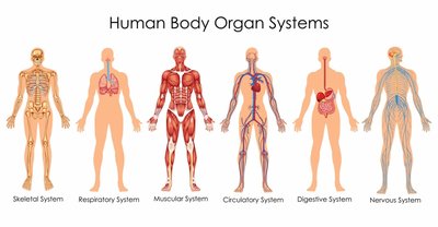
The human body is composed of several organ systems, each with specific functions:
Integumentary System
Skeletal System
Muscular System
Nervous System
Endocrine System
Cardiovascular System
Lymphatic/Immune System
Respiratory System
Digestive System
Urinary System
Reproductive System
Homeostasis: Maintaining Internal Balance
Definition and Importance
Homeostasis is the ability of the body to maintain relatively constant internal conditions, ensuring a stable environment for cellular processes. It involves dynamic equilibrium, where variables fluctuate within a normal range.
Feedback Mechanisms
Negative Feedback: The response reduces or eliminates the original stimulus, maintaining parameters (e.g., body temperature, heart rate, blood sugar) within a standard range. Most physiological processes use negative feedback.
Positive Feedback: The response amplifies the original stimulus, often promoting instability. Positive feedback is less common and usually associated with specific processes (e.g., childbirth, blood clotting).
Examples of Negative Feedback
Regulation of body temperature
Control of blood pressure
Regulation of blood glucose by insulin and glucagon
Examples of Positive Feedback
Hormonal control of parturition (childbirth)
Blood clotting cascade
Components of Homeostatic Systems
Stimulus: Change in the environment
Receptor: Detects the change
Control Center: Processes information and determines response
Effector: Carries out the response to restore balance
Standardized Terminology and Anatomical Position
Anatomical Position
The standard reference position for the body is:
Body erect
Feet on the floor, toes forward
Arms at sides, palms forward
Face forward
Directional Terms
Superior (Cranial): Above or toward the head
Inferior (Caudal): Below or toward the tail
Anterior (Ventral): Front or toward the belly
Posterior (Dorsal): Back or toward the spine
Medial: Toward the midline
Lateral: Away from the midline
Intermediate: Between medial and lateral
Proximal: Closer to the origin or trunk
Distal: Farther from the origin or trunk
Superficial (External): Toward the surface
Deep (Internal): Away from the surface
Planes of Section
Sagittal: Divides body into right and left portions
Midsagittal: Equal right and left portions
Parasagittal: Unequal right and left portions
Transverse (Horizontal): Divides body into superior and inferior portions
Frontal (Coronal): Divides body into anterior and posterior portions
Body Cavities and Membranes
Major Body Cavities
Dorsal Body Cavity: Contains the cranial cavity (brain) and vertebral cavity (spinal cord)
Ventral Body Cavity: Contains the thoracic and abdominopelvic cavities
The mediastinum is a central compartment in the thorax containing the heart and other structures.
Serous Membranes of the Ventral Cavity
Double-layered serous membranes line the ventral body cavity and cover organs:
Parietal layer: Lines the cavity wall
Visceral layer: Covers the organ
Serous fluid: Fills the space between layers, reducing friction
Three major serous membranes:
Pericardium: Surrounds the heart
Pleura: Surrounds the lungs
Peritoneum: Lines the abdominal cavity and covers abdominal organs
Special portions of the peritoneum include the greater omentum (attaches to the greater curvature of the stomach) and the lesser omentum (between the lesser curvature of the stomach and the liver).
Ascites is abnormal fluid accumulation in the peritoneal cavity, often due to cirrhosis.
Abdominopelvic Regions and Quadrants
The abdominopelvic cavity is divided for clinical and anatomical reference into regions and quadrants, aiding in the localization of organs and pain.
Additional info: This guide covers foundational concepts from the first chapter of a typical Anatomy & Physiology course, including anatomical terminology, organization, and homeostatic mechanisms. For further study, refer to subsequent chapters on cells, tissues, and organ systems.
