 뒤로
뒤로Introduction to Human Anatomy: Orientation, Body Regions, Planes, and Cavities
스터디 가이드 - 스마트 노트
 자료에 맞춘 맞춤형 노트, 핵심 정의, 예시, 맥락을 확장해 제공합니다.
Anatomical Position and Orientation
Standard Anatomical Position
The anatomical position is the universally accepted starting point for describing the human body’s location and orientation. This position ensures consistency in anatomical terminology and reference.
Body erect
Feet slightly apart
Palms facing forward
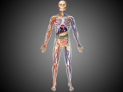
Importance: All directional terms (e.g., superior, inferior, anterior, posterior) are based on the body being in this position.
Regional Terms of the Human Body
Major Body Divisions
The body is divided into two main regions for anatomical reference:
Axial region: Includes the head, neck, and trunk.
Appendicular region: Includes the limbs (arms and legs).
Regional terms are used to designate specific areas within these divisions, aiding in precise location descriptions.
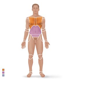
Examples of regional terms: Cephalic (head), Cervical (neck), Thoracic (chest), Brachial (arm), Femoral (thigh), etc.
Body Planes and Sections
Definition of Body Planes
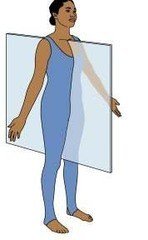
A plane is an imaginary flat surface that divides the body or an organ for anatomical study. Understanding these planes is essential for interpreting medical imaging and anatomical descriptions.
Sagittal plane: Divides the body into right and left parts.
Midsagittal (median) plane: Lies exactly on the midline.
Parasagittal plane: Offset from the midline.
Frontal (coronal) plane: Divides the body into anterior (front) and posterior (back) parts.
Transverse (horizontal) plane: Divides the body into superior (upper) and inferior (lower) parts; produces a cross-section.
Oblique section: Cuts made diagonally between horizontal and vertical planes.
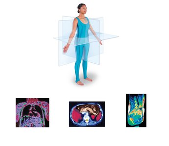
Example: Medical imaging (CT, MRI) often uses these planes to display internal structures.
Body Cavities and Membranes
Major Body Cavities
The body contains internal cavities that protect organs and allow for organ movement and expansion.
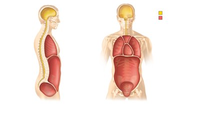
Dorsal body cavity: Protects the nervous system and has two subdivisions:
Cranial cavity: Encases the brain.
Vertebral cavity: Encases the spinal cord.
Ventral body cavity: Houses internal organs (viscera) and is divided by the diaphragm into:
Thoracic cavity: Contains heart and lungs; subdivided into pleural cavities (lungs), mediastinum (thoracic organs), and pericardial cavity (heart).
Abdominopelvic cavity: Contains abdominal cavity (digestive organs, spleen, liver) and pelvic cavity (urinary bladder, reproductive organs, rectum).
Serous Membranes (Serosa)
Serous membranes are thin, double-layered membranes that line the ventral body cavity and cover its organs, separated by a lubricating serous fluid.
Parietal serosa: Lines internal body walls.
Visceral serosa: Covers the internal organs.
Example: The pericardium surrounds the heart, with the parietal pericardium lining the cavity and the visceral pericardium covering the heart itself.
Abdominopelvic Quadrants
Clinical Division of the Abdomen
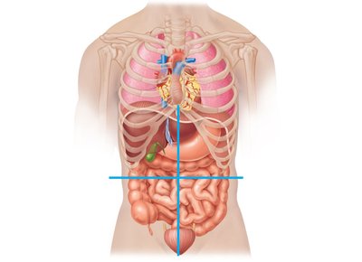
The abdominopelvic cavity is divided into four quadrants for clinical reference, aiding in diagnosis and communication among medical personnel.
Quadrant | Location |
|---|---|
Right Upper Quadrant (RUQ) | Upper right side |
Left Upper Quadrant (LUQ) | Upper left side |
Right Lower Quadrant (RLQ) | Lower right side |
Left Lower Quadrant (LLQ) | Lower left side |
Other Body Cavities
Additional Cavities
Besides the main dorsal and ventral cavities, the body contains several smaller cavities:
Oral and digestive cavities
Nasal cavity
Orbital cavities
Middle ear cavities
Synovial cavities (joint cavities)
These cavities serve specialized functions, such as housing sensory organs or facilitating joint movement.
