 뒤로
뒤로Introduction to Human Anatomy: Orientation, Planes, Cavities, and Organ Identification
스터디 가이드 - 스마트 노트
 자료에 맞춘 맞춤형 노트, 핵심 정의, 예시, 맥락을 확장해 제공합니다.
Orientation to the Human Body
Anatomical Position and Directional Terms
The anatomical position is a standardized stance used to describe locations and directions on the human body. Understanding anatomical directions is essential for accurately describing the position of structures.
Right vs. Left: Anatomical right and left refer to the subject's right and left, not the observer's.
Directional Terms:
Superior (Cranial): Toward the head or upper part of a structure.
Inferior (Caudal): Away from the head or toward the lower part of a structure.
Anterior (Ventral): Toward the front of the body.
Posterior (Dorsal): Toward the back of the body.
Body Planes and Sections
Major Anatomical Planes
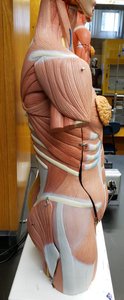
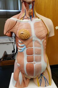
Body planes are imaginary lines used to divide the body into sections for anatomical study and medical imaging.
Sagittal Plane: Divides the body into right and left portions. The midsagittal (median) plane divides the body into equal right and left halves, while a parasagittal plane divides it unequally.
Frontal (Coronal) Plane: Divides the body into anterior (front) and posterior (back) portions.
Transverse (Horizontal) Plane: Divides the body into superior (upper) and inferior (lower) portions.
Body Cavities
Major Body Cavities and Their Contents
The human body contains several major cavities that house and protect vital organs.
Dorsal Body Cavity: Includes the cranial cavity (brain) and vertebral cavity (spinal cord).
Ventral Body Cavity: Subdivided into the thoracic cavity (heart and lungs) and abdominopelvic cavity (digestive, urinary, and reproductive organs).
Thoracic Cavity: Contains the pleural cavities (lungs) and the pericardial cavity (heart).
Abdominopelvic Cavity: Divided into the abdominal cavity (stomach, liver, intestines, etc.) and pelvic cavity (bladder, reproductive organs, rectum).
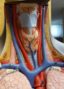
Organ Identification on Anatomical Models
Major Organs and Their Locations
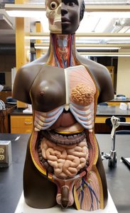
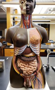
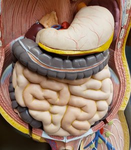
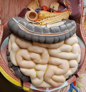
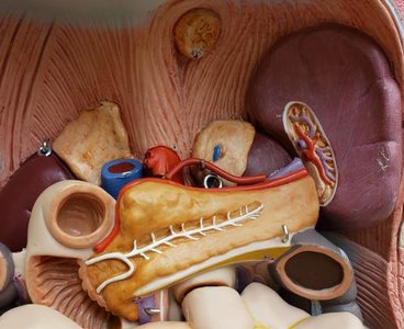
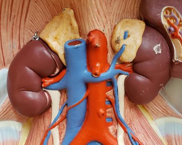
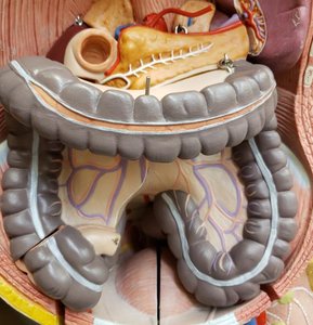
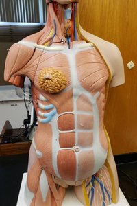
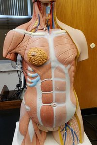
Being able to identify organs on anatomical models is a foundational skill in anatomy. The following images show various organs in situ and in isolation.
Heart: Located in the thoracic cavity, between the lungs.
Lungs: Paired organs flanking the heart in the thoracic cavity.
Liver: Large organ in the upper right abdomen, just below the diaphragm.
Stomach: Located in the upper left abdomen, below the liver.
Intestines: Occupy most of the abdominal cavity; small intestine is centrally located, surrounded by the large intestine.
Kidneys: Retroperitoneal organs located on the posterior abdominal wall.
Bladder: Located in the pelvic cavity, stores urine.
Reproductive Organs: Located in the pelvic cavity; male and female structures differ.
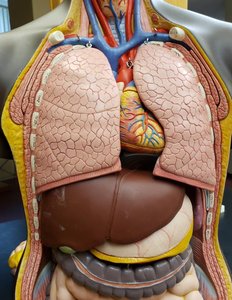
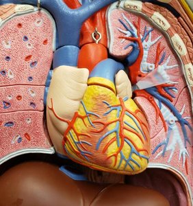
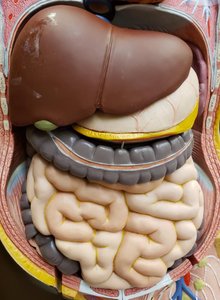
Abdominopelvic Quadrants and Regions
Divisions of the Abdominopelvic Cavity
The abdominopelvic cavity is divided into quadrants and regions to help locate organs and describe pain or pathology.
Quadrants: Right Upper Quadrant (RUQ), Left Upper Quadrant (LUQ), Right Lower Quadrant (RLQ), Left Lower Quadrant (LLQ).
Regions: Nine regions include the right/left hypochondriac, epigastric, right/left lumbar, umbilical, right/left iliac (inguinal), and hypogastric (pubic) regions.
Organ Structure and Function: Stomach Example
Stomach Anatomy and Function
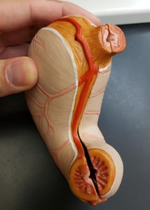
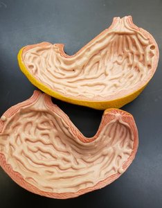
The stomach is a muscular organ involved in the mechanical and chemical breakdown of food. It has two main openings: the esophageal (cardiac) sphincter and the pyloric sphincter. The internal folds, called rugae, allow the stomach to expand after eating.
Esophageal Sphincter: Entry point for food from the esophagus.
Pyloric Sphincter: Exit point to the small intestine.
Rugae: Folds in the stomach lining that permit expansion.
Male Reproductive System and Associated Structures
Key Structures in the Male Pelvis
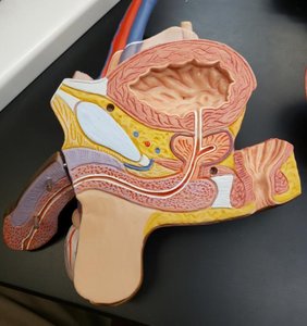
The male reproductive system includes the testes, ductus deferens, prostate, and penis. The bladder and rectum are also located in the pelvic cavity.
Testes: Produce sperm and testosterone.
Ductus Deferens: Transports sperm from the testes to the urethra.
Bladder: Stores urine before excretion.
Rectum: Terminal part of the large intestine, stores feces.
Comparative Anatomy: Rat Dissection
Identifying Organs in a Rat Model
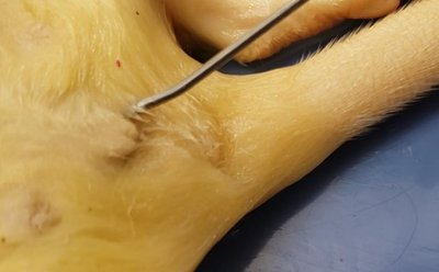
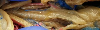
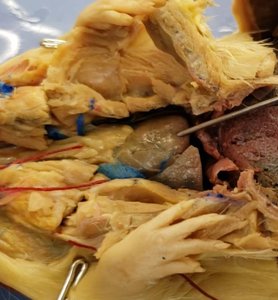
Dissection of a rat provides a comparative perspective on mammalian anatomy. Many organs are analogous to those in humans, including the liver, stomach, intestines, and kidneys.
Liver: Large, dark organ in the upper abdomen.
Stomach: Located just below the liver, often with visible rugae.
Intestines: Coiled structures occupying much of the abdominal cavity.
Kidneys: Bean-shaped organs located dorsally.
Summary Table: Major Body Cavities and Their Contents
Body Cavity | Major Organs |
|---|---|
Cranial | Brain |
Vertebral | Spinal cord |
Thoracic | Heart, lungs, esophagus, trachea |
Abdominal | Liver, stomach, spleen, intestines, kidneys |
Pelvic | Bladder, reproductive organs, rectum |
Additional info:
Understanding anatomical terminology and body orientation is foundational for all subsequent study in anatomy and physiology.
Comparative anatomy (e.g., rat dissection) reinforces the universality of organ systems among mammals.
