 뒤로
뒤로Introduction to Skeletal Tissues: Cartilage and Bone
스터디 가이드 - 스마트 노트
 자료에 맞춘 맞춤형 노트, 핵심 정의, 예시, 맥락을 확장해 제공합니다.
Introduction to Skeletal Tissues
The skeletal system is composed of specialized connective tissues that provide structure, support, and protection to the body. The two primary supporting connective tissues are cartilage and bone, each with unique properties and functions.
Classification of Connective Tissues
Connective tissues are broadly classified into three categories: connective tissue proper, fluid connective tissues, and supporting connective tissues. Cartilage and bone are the main supporting connective tissues, providing the framework for the body.
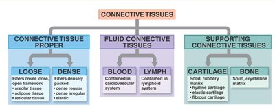
Cartilage
General Features of Cartilage
Cartilage is a resilient and smooth elastic tissue, a rubber-like padding that covers and protects the ends of long bones at the joints and is a structural component of the rib cage, ear, nose, bronchial tubes, and intervertebral discs.
Prefix "chondro-" refers to cartilage.
Cartilage contains few cells (chondrocytes) and an abundance of collagen fibers embedded in a ground substance called chondroitin sulfate, which may contain insoluble calcium salts.
All cartilage is avascular (lacks blood vessels).
Chondroblasts mature into chondrocytes, which reside in spaces called lacunae.
Suffixes in Skeletal Tissues
-blast: Indicates a cell that is growing or actively secreting extracellular matrix (e.g., chondroblast, osteoblast).
-clast: Indicates a cell that is breaking down matrix (e.g., osteoclast).
-cyte: Indicates a mature cell that maintains the tissue (e.g., chondrocyte, osteocyte).
Types of Cartilage
There are three main types of cartilage, each with distinct locations and functions:
Type | Main Function | Location | Histological Features |
|---|---|---|---|
Hyaline cartilage | Reduces friction at joints; keeps respiratory passages open | Ends of long bones, ribs, larynx, trachea | Chondrocytes in pairs, glassy appearance |
Elastic cartilage | Provides flexible framework | Auricle of ear, tip of nose, epiglottis | Netlike pattern of elastic fibers, large lacunae |
Fibrocartilage | Shock absorption, friction reduction, protection | Pubic symphysis, intervertebral discs | Rows of chondrocytes between collagen bundles |
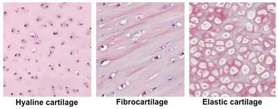
Hyaline Cartilage
Function: Reduces friction at joints, maintains open respiratory passages.
Location: Ends of long bones, ribs, larynx, trachea.
Features: Chondrocytes often found in pairs within lacunae; smooth, glassy appearance.
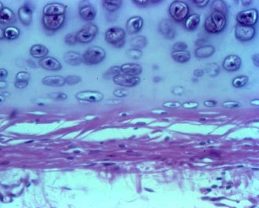
Elastic Cartilage
Function: Provides flexible support.
Location: Auricle of the ear, tip of the nose, epiglottis.
Features: Netlike pattern of dark elastic fibers; chondrocytes in large, closely packed lacunae.
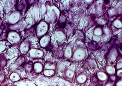
Fibrocartilage
Function: Shock absorption, friction reduction, protection of bones at joint surfaces.
Location: Pubic symphysis, intervertebral discs, menisci of knee.
Features: Numerous collagen fiber bundles; chondrocytes arranged in rows between fibers.
Bone (Osseous Tissue)
General Features of Bone
Bone is a rigid connective tissue that forms the skeleton. It provides support, protection, and enables movement. Bone tissue is highly vascularized and contains specialized cells and a mineralized matrix.
Prefix "osteo-" refers to bone.
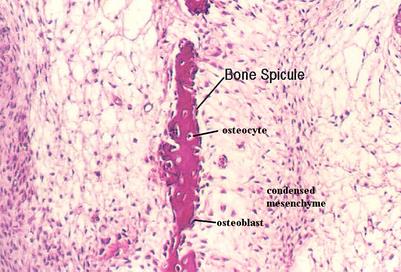
Osteoblasts secrete the organic component of bone matrix (osteoid), which is later mineralized into hydroxyapatite crystals and calcium salts.
Once surrounded by matrix, osteoblasts mature into osteocytes (reside in lacunae).
Osteoclasts are responsible for bone resorption (breakdown).
The periosteum is the outer covering; the endosteum lines the medullary cavity.
Types of Bone Tissue
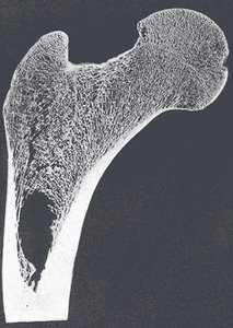
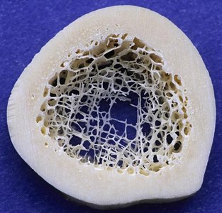
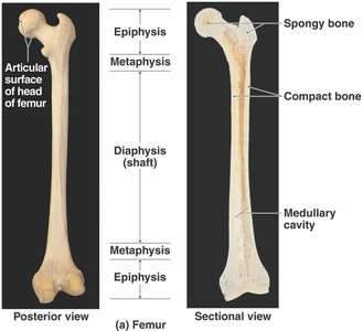
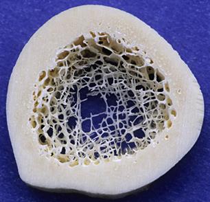
Spongy (cancellous/trabecular) bone: Composed of an open network of bony struts (trabeculae); directs forces and reduces weight.
Compact (cortical) bone: Dense, solid layers; provides strength and support.
Most bones contain both types.
Osteon: The Functional Unit of Compact Bone
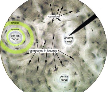
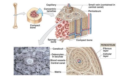
The osteon (Haversian system) is the basic structural unit of mature compact bone.
Osteocytes reside in lacunae between layers of matrix called lamellae (concentric and circumferential).
Central (Haversian) canal: Contains blood vessels and nerves.
Perforating (Volkmann's) canals: Run perpendicular to central canals, connecting blood supply.
Canaliculi: Tiny channels connecting lacunae, allowing communication and nutrient exchange.
Classification of Bones by Shape
Adult humans typically have 206 bones, classified by shape:
Long bones (e.g., femur, humerus)
Short bones (e.g., carpals, tarsals)
Flat bones (e.g., skull, sternum)
Irregular bones (e.g., vertebrae)
Sesamoid bones (e.g., patella)
Gross Anatomy of a Long Bone
Epiphyses: Ends of the bone, mostly spongy bone, covered with articular (hyaline) cartilage.
Diaphysis: Shaft, mostly compact bone, directs forces through the body.
Metaphysis: Region between diaphysis and epiphysis; in growing bones, contains the growth plate.
Medullary cavity: Central cavity containing yellow marrow (fat storage); red marrow (hematopoiesis) found in spongy bone of epiphyses and flat bones.
Bone Development (Ossification)
Types of Ossification
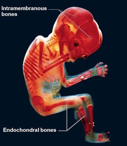
Intramembranous ossification: Bone develops directly from mesenchymal tissue (e.g., flat bones of skull).
Endochondral ossification: Bone forms by replacing a hyaline cartilage model (e.g., long bones).
Zones of Ossification (Epiphyseal Plate)
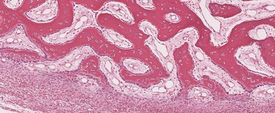
The growth plate (epiphyseal plate) is responsible for longitudinal bone growth and consists of several zones:
Resting (reserve) zone: Quiescent chondrocytes.
Proliferation zone: Rapidly dividing chondrocytes.
Hypertrophy zone: Chondrocytes enlarge and mature.
Calcification zone: Chondrocytes die, matrix calcifies, and is replaced by bone tissue.
Mnemonic: Rats Prefer Houses with Cheese (Resting, Proliferation, Hypertrophy, Calcification).
Summary Table: Cartilage vs. Bone
Feature | Cartilage | Bone |
|---|---|---|
Cells | Chondrocytes in lacunae | Osteocytes in lacunae |
Matrix | Chondroitin sulfate, collagen/elastic fibers | Osteoid, hydroxyapatite, collagen fibers |
Vascularity | Avascular | Highly vascular |
Growth | Appositional and interstitial | Mainly appositional |
Types | Hyaline, elastic, fibrocartilage | Compact, spongy |
Example: The femur is a long bone that develops via endochondral ossification, contains both compact and spongy bone, and has articular cartilage at its epiphyses.
Additional info: Bone remodeling is a lifelong process involving osteoblasts (formation) and osteoclasts (resorption), essential for calcium homeostasis and repair.
