 뒤로
뒤로Lab 1: Organ Survey and Histology Part 1 – Epithelial and Muscle Tissues
스터디 가이드 - 스마트 노트
 자료에 맞춘 맞춤형 노트, 핵심 정의, 예시, 맥락을 확장해 제공합니다.
Major Organs and Organ Systems
Introduction to Major Organs
Understanding the location and function of major organs is foundational in anatomy and physiology. Each organ belongs to a specific organ system, contributing to the body's overall function and homeostasis.
Nervous System: Brain
Cardiovascular System: Heart
Lymphatic System: Spleen
Respiratory System: Lungs
Digestive System: Liver, Gallbladder, Stomach, Pancreas, Small Intestine, Large Intestine
Urinary System: Kidneys, Urinary Bladder
Endocrine System: Adrenal Glands, Thyroid Gland
Example: The heart is the central organ of the cardiovascular system, responsible for pumping blood throughout the body.
Histology: The Study of Tissues
Introduction to Histology
Histology is the science of studying the microscopic structure of tissues. All tissues in the human body are classified into four general types: epithelial, muscle, connective, and nervous tissue. This lab focuses on epithelial and muscle tissues.
Epithelial Tissue: Covers body surfaces and lines cavities.
Muscle Tissue: Responsible for movement.
Epithelial Tissue
General Characteristics of Epithelial Tissue
Epithelial tissues cover surfaces exposed to the environment and line hollow organs and body cavities. The cells are tightly packed with minimal extracellular space and are anchored to underlying tissues by a basement membrane, which consists of the basal lamina and reticular lamina. The apical surface faces the environment or lumen, while the basal surface attaches to the basement membrane.
Classification Criteria: Cell shape and number of layers.
Cell Shapes: Squamous (flat), Cuboidal (cube-shaped), Columnar (tall).
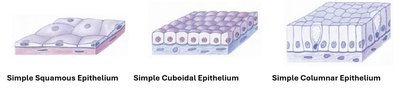
Example: The lining of blood vessels is composed of simple squamous epithelium, allowing efficient diffusion.
Classification by Layers
Epithelial tissues are further classified by the number of cell layers:
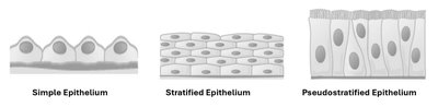
Simple Epithelium: Single layer; all cells touch the basement membrane.
Stratified Epithelium: Multiple layers; named for the shape of the apical cells.
Pseudostratified Epithelium: Appears multilayered but all cells contact the basement membrane.
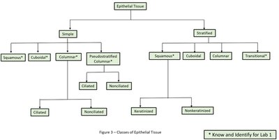
Classification Overview
The following chart summarizes the main classes of epithelial tissue, including those required for Lab 1:
Types of Epithelial Tissue
Simple Squamous Epithelium
Composed of a single layer of thin, flat cells, this tissue allows for rapid diffusion, osmosis, filtration, and secretion. Special names include endothelium (lining heart and blood vessels) and mesothelium (lining body cavities).
Locations: Alveoli, glomerular capsule, lining of body cavities, heart chambers, blood vessels
Simple Cuboidal Epithelium
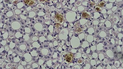
Consists of a single layer of cube-shaped cells, often involved in secretion and absorption, with limited protective function.
Locations: Thyroid gland, lining kidney tubules
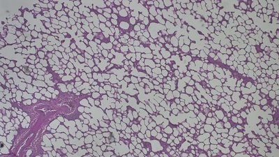
Simple Columnar Epithelium
Composed of a single layer of tall, column-like cells. May have cilia (for movement) or microvilli (for increased surface area). Functions in absorption and secretion.
Locations: Lining of the stomach, gallbladder, small intestine, large intestine
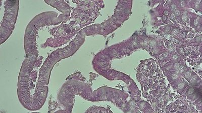
Pseudostratified Columnar Epithelium
Appears stratified due to varying cell heights, but all cells contact the basement membrane. Contains goblet cells (mucus production) and cilia (movement of substances).
Locations: Lining of the trachea, nasal cavities
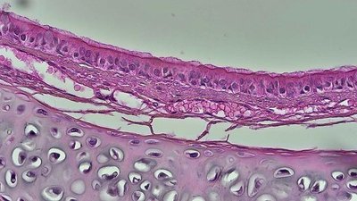
Stratified Squamous Epithelium
Multiple layers of cells with squamous cells at the apical surface. Provides protection in areas subject to abrasion and mechanical stress.
Locations: Epidermis, lining of esophagus, anus, rectum, vagina
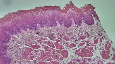
Transitional Epithelium
Multiple layers with dome-shaped apical cells. Specialized to stretch and recoil without damage.
Locations: Lining of ureters, urinary bladder
Muscle Tissue
Overview of Muscle Tissue Types
Muscle tissue is specialized for contraction and movement. There are three types:
Skeletal Muscle: Voluntary, striated, multinucleate, cylindrical cells. Found in skeletal muscles.
Cardiac Muscle: Involuntary, striated, single nucleus, branched cells, intercalated discs. Found in the heart.
Smooth Muscle: Involuntary, non-striated, single nucleus, tapered cells. Found in walls of blood vessels and hollow organs.
Lab 1 Study Checklist
Define all bold terms from the lab manual.
Identify the major organs listed.
Classify each tissue as muscle or epithelium.
Identify the 3 muscle tissues and 6 epithelial tissues by their complete names.
Label bolded structures for each tissue.
Give one detailed body location for each tissue, as listed in the manual.
Use only lab manual definitions and terms.
Summary Table: Epithelial Tissues for Lab 1
Tissue Name | Number of Layers | Cell Shape | Key Locations | Special Features |
|---|---|---|---|---|
Simple Squamous Epithelium | 1 | Flat | Alveoli, blood vessels, body cavities | Rapid diffusion, filtration |
Simple Cuboidal Epithelium | 1 | Cube-shaped | Kidney tubules, thyroid gland | Secretion, absorption |
Simple Columnar Epithelium | 1 | Tall, column-like | Stomach, intestines, gallbladder | Absorption, secretion, may have cilia/microvilli |
Pseudostratified Columnar Epithelium | 1 (appears multilayered) | Columnar | Trachea, nasal cavities | Cilia, goblet cells (mucus) |
Stratified Squamous Epithelium | Multiple | Flat at surface | Epidermis, esophagus, anus, vagina | Protection from abrasion |
Transitional Epithelium | Multiple | Dome-shaped at surface | Urinary bladder, ureters | Stretch and recoil |
