 뒤로
뒤로Lab 3 : Major Systemic Arteries, Veins, and Blood Pressure
스터디 가이드 - 스마트 노트
 자료에 맞춘 맞춤형 노트, 핵심 정의, 예시, 맥락을 확장해 제공합니다.
Major Systemic Arteries, Veins, and Blood Pressure
Overview of the Circulatory System
The circulatory system is responsible for transporting blood, nutrients, gases, and wastes throughout the body. It consists of the heart, blood vessels (arteries, veins, and capillaries), and blood. This guide focuses on the major systemic arteries and veins, as well as the principles of blood pressure measurement.
Systemic Arteries
Major Arteries of the Body
Arteries carry oxygenated blood away from the heart to the tissues of the body. The largest artery is the aorta, which branches into major arteries serving the head, neck, upper extremities, thorax, abdomen, pelvis, and lower extremities.
Aorta: Divided into ascending aorta, aortic arch, and descending aorta (thoracic and abdominal parts).
Brachiocephalic artery (trunk): First major branch of the aortic arch, giving rise to the right subclavian and right common carotid arteries.
Subclavian arteries: Supply the upper limbs and parts of the thorax.
Common carotid arteries: Each divides into internal and external carotid arteries, supplying the head and neck.
Vertebral arteries: Supply the posterior part of the brain.
Axillary, brachial, radial, and ulnar arteries: Supply the upper extremities.
Descending aorta branches: Include esophageal, intercostal, phrenic, celiac trunk (with splenic, hepatic, and gastric branches), superior and inferior mesenteric, renal, gonadal, and lumbar arteries.
Common, internal, and external iliac arteries: Supply the pelvis and lower limbs.
Femoral, popliteal, tibial, dorsalis pedis, and arcuate arteries: Supply the lower extremities.


Circle of Willis
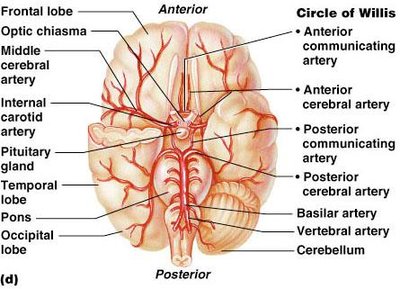
The Circle of Willis is a circular arterial anastomosis located at the base of the brain. It encircles the pituitary gland and unites the brain’s anterior and posterior blood supplies. This structure helps equalize blood pressure in the brain and provides alternate routes for blood flow if a major artery is blocked.
Formed by the anterior cerebral, anterior communicating, internal carotid, posterior cerebral, and posterior communicating arteries.
Fed by the basilar artery (from vertebral arteries) and internal carotid arteries.

Systemic Veins
Major Veins of the Body
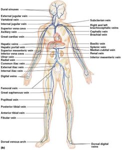
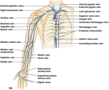
Veins return deoxygenated blood from the body to the heart. The largest veins are the superior and inferior vena cava, which drain the upper and lower parts of the body, respectively.
Superior vena cava: Drains blood from the head, neck, upper limbs, and thorax.
Inferior vena cava: Drains blood from the abdomen, pelvis, and lower limbs.
Jugular veins (internal and external): Drain the head and neck.
Brachiocephalic veins: Formed by the union of the subclavian and internal jugular veins.
Subclavian, axillary, brachial, radial, and ulnar veins: Drain the upper extremities.
Common, internal, and external iliac veins: Drain the pelvis and lower limbs.
Femoral, great saphenous, popliteal, tibial, and fibular veins: Drain the lower extremities.

Hepatic Portal System
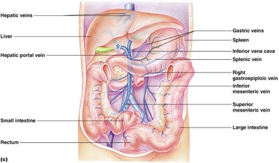
The hepatic portal system is a specialized venous system that drains nutrient-rich blood from the digestive organs and routes it through the liver before returning to the heart. This allows the liver to detoxify substances and regulate nutrient levels.
Major veins: hepatic portal vein, superior mesenteric vein, splenic vein, gastric veins, right gastroepiploic vein, and hepatic veins.
Liver cells remove excess glucose (stored as glycogen) and store some vitamins.

Blood Pressure
Measuring Blood Pressure
Blood pressure is the force exerted by circulating blood on the walls of blood vessels, typically measured in the brachial artery. It is recorded as two numbers: systolic (during heart contraction) and diastolic (during heart relaxation).
Measured using a sphygmomanometer and stethoscope (auscultation method).
Normal blood pressure: < 120/80 mmHg.
Categories of blood pressure:
Category | Systolic (mmHg) | Diastolic (mmHg) |
|---|---|---|
Optimal | < 120 | < 80 |
Prehypertensive | 120–139 | 80–89 |
Stage 1 Hypertension | 140–159 | 90–99 |
Stage 2 Hypertension | 160–179 | 100–109 |
Stage 3 Hypertension | 180–209 | 110–119 |
Stage 4 Hypertension | ≥ 210 | ≥ 120 |

Pulse and Pulse Points
The pulse is the rhythmic expansion and recoil of arteries as blood is pumped by the heart. Pulse points are locations where the pulse can be easily felt by compressing an artery against a firm structure.
Common pulse points: temporal, facial, carotid, brachial, radial, femoral, popliteal, posterior tibial, dorsalis pedis arteries.
Summary Table: Major Arteries and Veins
Region | Major Arteries | Major Veins |
|---|---|---|
Head & Neck | Common carotid, internal/external carotid, vertebral | Internal/external jugular, vertebral, brachiocephalic |
Upper Extremities | Subclavian, axillary, brachial, radial, ulnar | Subclavian, axillary, brachial, cephalic, basilic, median cubital |
Thorax | Aorta, intercostal arteries | Superior vena cava, azygos vein |
Abdomen | Abdominal aorta, celiac trunk, mesenteric, renal, gonadal | Inferior vena cava, hepatic, renal, gonadal |
Pelvis & Lower Extremities | Common iliac, internal/external iliac, femoral, popliteal, tibial | Common iliac, internal/external iliac, femoral, great saphenous, popliteal, tibial |
Additional info:
Arteries are typically deeper and have thicker walls than veins due to higher pressure.
Veins have valves to prevent backflow, especially in the limbs.
The hepatic portal system is unique in that it connects two capillary beds (digestive organs and liver) before returning blood to the heart.
