 뒤로
뒤로Male Reproductive System: Structure, Function, and Spermatogenesis
스터디 가이드 - 스마트 노트
 자료에 맞춘 맞춤형 노트, 핵심 정의, 예시, 맥락을 확장해 제공합니다.
Male Reproductive System
Overview and Objectives
The male reproductive system is responsible for the production, maturation, and delivery of spermatozoa, as well as the synthesis of male sex hormones. Understanding its anatomy and physiology is essential for comprehending human reproduction and related clinical conditions.
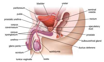
Organs: Includes main (testes, penis) and accessory (prostate gland, seminal vesicles, bulbourethral glands) structures.
Functions: Sperm production, hormone synthesis, sperm transport, and clinical relevance.
Anatomical Components
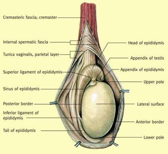
Testes: Paired ovoid organs located in the scrotum, responsible for spermatogenesis and steroidogenesis.
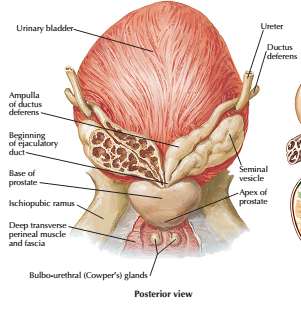
Genital Duct System: Includes epididymis, ductus deferens, ejaculatory ducts, and urethra.
Accessory Sex Glands: Prostate gland, seminal vesicles, bulbourethral glands.
Penis: External organ for copulation and urine excretion.
Testis: Structure and Function
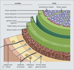
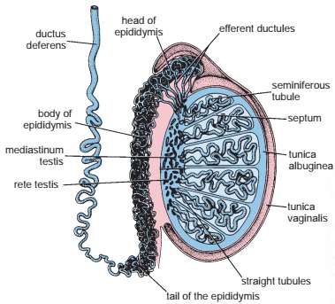
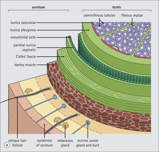
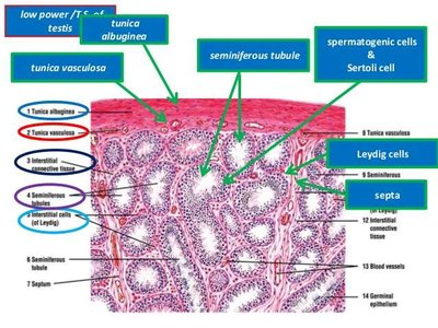
The testis is the primary male reproductive organ, enclosed by the tunica albuginea and divided into lobules containing seminiferous tubules and interstitial cells.
Tunica albuginea: Dense connective tissue capsule surrounding the testis.
Tunica vasculosa: Loose connective tissue layer beneath the tunica albuginea.
Mediastinum testis: Thickened posterior region projecting inward.
Lobules: Each contains 1-4 seminiferous tubules and Leydig cells.
Seminiferous Tubules
Seminiferous tubules are the site of sperm production and are surrounded by peritubular tissue containing myoid cells.
Seminiferous epithelium: Stratified epithelium containing spermatogenic and Sertoli cells.
Tunica propria: Multilayered connective tissue with myoid cells.
Functions: Spermatogenesis and steroidogenesis.
Cell Types in Seminiferous Tubules
The seminiferous epithelium contains two main cell populations: spermatogenic (germ) cells and Sertoli (supporting) cells.
Spermatogenic cells: Undergo regular replication and differentiation to form mature sperm.
Sertoli cells: Support, nourish, and protect developing sperm; form the blood-testis barrier.
Spermatogenesis
Phases of Spermatogenesis
Spermatogenesis is the process by which sperm are produced, initiated by gonadotrophins at puberty and divided into three phases:
Spermatocytogenesis: Differentiation of spermatogonia into primary spermatocytes (mitosis).
Meiosis: Reduction division to form haploid spermatids from primary spermatocytes.
Spermiogenesis: Transformation of spermatids into spermatozoa (cytodifferentiation).
Key formula:
$\text{Spermatogonia} \xrightarrow{\text{Mitosis}} \text{Primary Spermatocytes} \xrightarrow{\text{Meiosis I}} \text{Secondary Spermatocytes} \xrightarrow{\text{Meiosis II}} \text{Spermatids} \xrightarrow{\text{Spermiogenesis}} \text{Spermatozoa}$
Spermatogonia Subtypes
There are three subtypes of spermatogonia in humans:
Type A (dark): Reserve stem cells, mitotically inactive.
Type A (pale): Active stem cells, undergo mitosis to produce Type B cells.
Type B: Undergo growth and become primary spermatocytes.
Spermiogenesis: Stages
Spermiogenesis is the final stage of sperm development, involving cytodifferentiation and the formation of mature spermatozoa.
Golgi phase: Formation of acrosomal granule.
Cap phase: Expansion of acrosomal vesicle over nucleus.
Acrosomal phase: Nuclear condensation and mitochondrial aggregation.
Maturation phase: Loss of excess cytoplasm, release of spermatozoa into lumen.
Sertoli Cells: Functions
Sertoli cells are essential for supporting spermatogenesis and maintaining the blood-testis barrier.
Support and nourish developing sperm.
Phagocytose excess cytoplasm.
Release mature sperm into lumen.
Synthesize androgen-binding protein (ABP).
Secrete inhibin and anti-müllerian hormone.
Form blood-testis barrier.
Blood-Testis Barrier
The blood-testis barrier is formed by tight junctions between Sertoli cells, dividing the seminiferous tubule into basal and adluminal compartments. It protects developing sperm from autoimmune reactions.
Basal compartment: Contains spermatogonia and early primary spermatocytes.
Adluminal compartment: Contains more mature spermatocytes and spermatids.
Clinical relevance: Failure of isolation can lead to production of sperm-specific antibodies, causing infertility.
Genital Ducts
Intratesticular and Extratesticular Ducts
The genital duct system transports sperm from the testis to the exterior.
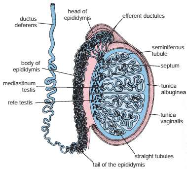
Intratesticular ducts: Straight tubules, rete testis, proximal efferent ductules.
Extratesticular ducts: Distal efferent ductules, ductus epididymis, ductus deferens, ejaculatory ducts, urethra.
Epididymis
The epididymis is a coiled tube where sperm mature and are stored.
Epithelium: Pseudostratified columnar with stereocilia.
Functions: Storage, maturation, absorption of fluid, phagocytosis of residual cytoplasm.
Ductus (Vas) Deferens
The ductus deferens is a muscular tube that transports sperm from the epididymis to the ejaculatory duct.
Length: 30-45 cm.
Epithelium: Pseudostratified columnar with stereocilia.
Muscularis: Three layers (inner longitudinal, middle circular, outer longitudinal).
Urethra
The male urethra serves as the terminal duct for both urinary and genital systems, with three segments:
Prostatic urethra: Lined with transitional epithelium.
Membranous urethra: Stratified or pseudostratified columnar epithelium.
Penile (spongy) urethra: Stratified or pseudostratified columnar epithelium, distal end lined with stratified squamous epithelium.
Accessory Glands
Prostate Gland
The prostate gland produces a clear, slightly alkaline fluid rich in citric acid, prostatic acid phosphatase, amylase, fibrinolysin, and prostate-specific antigen (PSA).
Functions: Liquefies semen, activates sperm, neutralizes acidity.
Clinical relevance: Prostate enlargement can cause urinary symptoms and complications.
Bulbourethral (Cowper's) Glands
These glands produce a clear, mucous-like secretion that lubricates the penile urethra prior to emission of semen.
Secretions: Contain galactose, galactosamine, galacturonic acid, sialic acid, methylpentose.
Seminal Vesicles
Seminal vesicles produce a whitish yellow viscous fluid containing fructose, fibrinogen, amino acids, ascorbic acid, and prostaglandins.
Functions: Provides energy for sperm, contributes to semen volume.
Penis
Structure
The penis contains three erectile tissues: two corpora cavernosa and one corpus spongiosum, which surrounds the penile urethra.
Tunica albuginea: Fibrous sheath surrounding erectile tissues.
Glans penis: Dilated distal end of corpus spongiosum.
Glands of Littre: Mucus-secreting glands throughout penile urethra.
Semen/Seminal Fluid
Semen contains sperm and secretory products from the testis, epididymis, ductus deferens, prostate, seminal vesicles, and bulbourethral glands.
Functions: Transport medium for sperm, neutralizes acidity, liquefies coagulated semen, activates sperm after ejaculation.
Clinical Correlations
Blood-Testis Barrier and Immunology
The blood-testis barrier isolates spermatogenic cells from the immune system. Failure of isolation can lead to production of sperm-specific antibodies, causing infertility.
After vasectomy: Sperm-specific antibodies may be produced, leading to agglutination and infertility.
Prostate enlargement: Can cause urinary retention, infection, and kidney failure.
Summary Table: Male Reproductive System Components
Component | Structure | Function |
|---|---|---|
Testis | Tunica albuginea, lobules, seminiferous tubules | Spermatogenesis, steroidogenesis |
Epididymis | Coiled tube, pseudostratified epithelium | Sperm maturation and storage |
Ductus deferens | Muscular tube, three muscle layers | Sperm transport |
Prostate gland | Fibromuscular, glandular tissue | Seminal fluid production |
Seminal vesicles | Highly folded mucosa | Seminal fluid production |
Bulbourethral glands | Mucous glands | Lubrication |
Penis | Corpora cavernosa, corpus spongiosum | Copulation, urine excretion |
